Comparison of survival benefit and safety profile between lenvatinib and donafenib as conversion therapy in patients with hepatocellular carcinoma 

In a retrospective comparative cohort study, Hou et al. 1) published in the American Journal of Translational Research, the authors—affiliated with the Department of Oncology, Department of Gynaecology and Obstetrics, and Department of Neurosurgery at Shijiazhuang People’s Hospital (Hebei, China), as well as Beijing Water Conservancy Hospital—compared the survival benefit and safety profile of lenvatinib versus donafenib as conversion therapy in patients with hepatocellular carcinoma (HCC) at China National Liver Cancer (CNLC) stages I–III.


Lenvatinib demonstrated significantly superior survival outcomes—both in overall survival and progression-free survival—compared to donafenib. It also showed better tolerability, with fewer grade ≥3 adverse events.


❌ 1. Study Design: Retrospective = Weak Evidence

This is yet another retrospective single-center analysis, plagued by inherent biases—selection, reporting, and confounding—that no amount of statistical massaging can resolve. No randomization, no blinding, and no control for treatment timing or physician discretion. In oncology, where treatment nuances matter, such designs should be considered hypothesis-generating at best, not guidance for clinical practice.

Read more

Temporal trends and risk factors associated with stroke mortality among cancer patients

In a retrospective cohort study published in the Journal of Clinical Neuroscience, Ahmed et al. with Cleveland Clinic Cerebrovascular Center, West Virginia University participation 1) analyzed data from over 5.9 million patients diagnosed with a first primary cancer, based on the SEER database (2000–2020). The study aimed to quantify the risk of stroke-related death (SD) in cancer patients and to identify temporal trends and associated clinical and demographic risk factors. Stroke-related mortality (SD) among cancer patients has significantly declined over the past two decades across all cancer types and both sexes. However, older age, non-white race, male sex, and specific cancer types—notably nervous system, respiratory, and head and neck cancers—are associated with a higher risk of stroke death. Conversely, patients receiving chemotherapy or radiotherapy had a lower risk of SD compared to those who received no treatment.


⚠️ Fatal Methodological Flaws

No Clinical Stroke Classification

The authors report on “stroke mortality” without differentiating ischemic vs. hemorrhagic strokes, nor providing stroke etiology or timing relative to cancer diagnosis or cancer treatment—rendering any mechanistic or preventative inference purely speculative.

Read more

Engineering of CD63 Enables Selective Extracellular Vesicle Cargo Loading and Enhanced Payload Delivery.

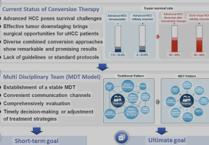
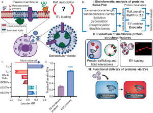
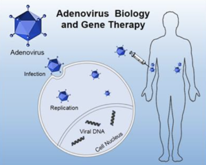
In a preclinical experimental study, Obuchi et al. (2025)—with contributions from the Department of Neurosurgery at Leiden University Medical Center—engineered extracellular vesicles for selective cargo loading and enhanced functional delivery, using a modified CD63 scaffold and VSV-G fusion, with in vivo validation in mouse brain models. 1)


🚫 1. Rebranding Complexity as Innovation The authors tout a modular EV engineering system using CD63, mCherry, FLAG-tags, nanobody fusions, and VSV-G. But this is not scientific ingenuity—it’s molecular bricolage. Each component is repurposed from older literature and glued together without real conceptual novelty. The result? A bloated acronym soup with more moving parts than scientific value.

⚠️ What’s pitched as a breakthrough is closer to a tech demo in search of a clinical rationale.

❌ 2. Absence of Disease-Relevant Application Despite name-dropping CRISPR, Cre, and Cas9, no disease context is addressed. No glioma model. No neurodegenerative target. No proof that the cargo accomplishes anything biologically meaningful in the recipient tissue. The mouse brain “validation” is just a fluorescent readout, not a therapeutic outcome.

The cargo arrives, but so what? This is payload delivery without a payload purpose.

🧪 3. Methodological Blind Spots No quantification of EV heterogeneity or functional subpopulations.

No rigorous comparison with alternative delivery systems (e.g., AAVs, lipid nanoparticles).

No evidence of endosomal escape for actual cytoplasmic/nuclear action.

No dose-response curves, toxicity profiling, or repeatability metrics.

This is a biotech prototype, not a therapy-in-the-making.

🔥 4. VSV-G: The Short-Term High, Long-Term Problem The use of VSV-G, a viral fusogen with broad tropism and high immunogenicity, is particularly careless. While it boosts in vitro uptake and helps “sell” delivery efficiency, it introduces a critical translational liability: poor specificity, potential immune activation, and unsuitability for clinical use.

❝Putting VSV-G on EVs is like installing a rocket engine on a paper boat—it moves faster, but it’s doomed to burn out or sink.❞

🧱 5. Structural Inefficiency and Complexity The system requires:

Engineering CD63 with dual tags

Fusing cargo to a nanobody

FLAG-based purification

VSV-G pseudotyping

TEV protease cleavage

This is logistically unscalable for clinical or industrial production and riddled with points of failure. The more components you bolt on, the more it resembles a lab curiosity, not a deliverable platform.

📉 6. Journal Inflation and Institutional Complacency The publication in J Extracell Vesicles is not a mark of impact, but rather a reflection of how EV journals have drifted into translational cosplay, applauding synthetic elegance over clinical relevance. The heavyweight affiliations (MGH, Harvard, etc.) likely ensured acceptance despite the absence of therapeutic depth or mechanistic rigor.

🧠 Conclusion: A study more interested in showing what’s technically possible than what’s biologically meaningful. It trades therapeutic relevance for engineering flair, while ignoring the hard questions of targeting, safetyscalability, and necessity.

This is not a step toward Evidence-based medicine—it’s a flashy side road to nowhere.


1)

Obuchi W, Zargani-Piccardi A, Leandro K, Rufino-Ramos D, Di Lanni E, Frederick DM, Maalouf K, Nieland L, Xiao T, Repiton P, Vaine CA, Kleinstiver BP, Bragg DC, Lee H, Miller MA, Breakefield XO, Breyne K. Engineering of CD63 Enables Selective Extracellular Vesicle Cargo Loading and Enhanced Payload Delivery. J Extracell Vesicles. 2025 Jun;14(6):e70094. doi: 10.1002/jev2.70094. PMID: 40527733.

Epithelioid angiosarcoma of the cervical spine: A case report.

Nan et al. 1) describe a rare case of epithelioid angiosarcoma (EA) involving the cervical spine, presenting with pathological fracture and kyphotic deformity, and document the surgical and adjuvant management as well as the clinical outcome in the World Journal of Clinical Cases.

1. Predictable Yet Pointless

The authors claim novelty by describing a rare anatomical presentation of EA. However, this degenerates into a predictable narrative with no new pathophysiological insights, no hypothesis generated, and no clinical paradigm challenged. It is the kind of “rare case” that proliferates in low-barrier journals precisely *because* it demands no intellectual risk.

2. Zero Diagnostic Value

The authors bypass the opportunity to deepen our understanding of the radiological-morphological signature of EA in the spine. No comparative imaging, no differential diagnostic flowchart, no histopathological discussion beyond standard CD31/CD34 immunostaining. If this case had been published in 1995, it would be equally uninformative.

3. Therapeutic Confusion Disguised as Aggressiveness

Two major spine surgeries (posterior decompression + anterior corpectomy) followed by immediate radiotherapy in a moribund patient demonstrate therapeutic overreach without oncological strategy. There is no discussion on multidisciplinary planning, palliative thresholds, or whether delaying surgery or avoiding the second procedure might have prevented ARDS. The reader is left with the impression of a surgical reflex, not an evidence-based decision.

4. No Discussion of Differential Diagnosis or Biomarkers

In a tumor type notorious for being misdiagnosed as metastasischordoma, or sarcoma NOS, the absence of a differential diagnostic framework or advanced markers (ERG, FLI1, HHV-8, etc.) is alarming. Histological laziness cloaked in “rare disease” rhetoric.

5. Outcome Reporting: Conveniently Truncated

The patient dies 3 weeks after surgery, yet the discussion fails to draw any causal or cautionary link between the interventions and the fatal ARDS. No autopsy data, no postmortem imaging, no pulmonary workup. This omission sterilizes the clinical narrative, reducing it to anecdote.

6. Ethically Murky

The case implicitly raises an ethical dilemma—should maximal surgery be performed in aggressive, terminal tumors without demonstrated systemic control? Yet the authors shy away from even mentioning this, let alone framing it for academic discussion.

7. Journal Choice Reflects the Paper’s Weakness

Published in a journal known for minimal peer review stringency, the article offers no citations of recent molecular or targeted therapy advances, no engagement with broader oncological guidelines, and no rationale for the treatment decisions beyond procedural listing.

  • Histological laziness: Failing to provide in-depth pathology discussion beyond CD31/CD34 and H&E staining in vascular tumors.
  • Surgical reflex: The tendency to operate based on mechanical findings (compression, fracture) without integrating prognosis or systemic disease behavior.
  • Ethical sterilization: Avoiding uncomfortable questions about futility, risk-benefit tradeoffs, and overtreatment in end-stage patients.
  • Postmortem evasion: Reporting a perioperative death without diagnostic closure (autopsy, imaging, or medical reflection).

This case report is an example of procedural reporting devoid of scientific merit, clinical reflection, or ethical introspection. It contributes nothing to the understanding of EA, its diagnosis, biology, or management—beyond reiterating its rarity. In its current form, it is neither hypothesis-generating nor practice-changing, and serves as a cautionary tale on how not to write a case report.

  • Include comparative radiology with metastatic disease and primary bone tumors.
  • Provide autopsy findings or detailed explanation of respiratory decline.
  • Discuss therapeutic alternatives (e.g., single-stage surgery, biopsy + RT, palliative care).
  • Frame the case within an oncological decision-making algorithm.

1)

Nan YH, Chiu CD, Chen WL, Chen LC, Chen CC, Cho DY, Guo JH. Epithelioid angiosarcoma of the cervical spine: A case report. World J Clin Cases. 2025 Jun 16;13(17):101593. doi: 10.12998/wjcc.v13.i17.101593. PMID: 40524767; PMCID: PMC11866273.

Histological Classifier of Radiosensitivity to Spine Stereotactic Body Radiation Therapy

In a Retrospective Cohort Study Jackson et al. 1) from the Memorial Sloan Kettering Cancer Center, New York, concluded that 30 Gy in 3 fractions is the preferred SBRT regimen for spinal metastases, even in radiosensitive tumors, because it offers better local control than 27 Gy with a similar risk of vertebral fracture requiring treatment.

Additionally:

Tumor histology strongly influences radiosensitivity — prostate and breast (Class A) respond best; GI and liver tumors (Class C) have higher failure rates.

For high-grade epidural compression (ESCC 2–3) in Class B–C tumors, separation surgery + SBRT may improve outcomes over SBRT alone.


* The study assigns biological significance to histology classes (A–C) without molecular stratification or genomic profiling — a gross simplification that ignores intratumoral heterogeneity and microenvironmental factors. * Retrospective design with non-randomized treatment allocation allows substantial selection bias (e.g., healthier patients might be more likely to receive 30 Gy). * No formal validation cohort — the classifier is proposed based on internal data, without prospective or external validation.

* “Histological classifier of radiosensitivity” suggests a predictive tool, yet the study lacks any predictive modeling or decision support framework. The term is more rhetorical than scientific. * The study mixes observational epidemiology with causal language, implying therapeutic superiority without proper adjustment for confounders.

* Treatment practices evolved over the 9-year window. Earlier patients may have been treated with less advanced planning, different immobilization, or different imaging standards — contaminating outcome comparisons.

* No adjustment for institutional learning curve, planning margins, spinal cord tolerance constraints, or radiologist/radiation oncologist variability. * The impact of systemic therapy (e.g., concurrent immunotherapytargeted therapy) is not accounted for — a major omission in modern oncologic outcomes.

* Vertebral Compression Fracture rates are reported, but the distinction between radiologic and clinically significant fractures is vague. Moreover, attributing cause solely to dose without biomechanical modeling is speculative. * The apparent increase in overall VCF with 30 Gy, though dismissed as statistically irrelevant, raises safety concerns insufficiently explored.

* The “benefit” of separation surgery in Epidural Spinal Cord Compression (ESCC) 2–3 lesions is statistically non-significant (p = 0.051) and based on a small subgroup (n=261) — yet it is discussed as if near-clinical truth.

This study sells the illusion of a refined, histology-based SBRT dosing paradigm, but offers little more than retrospective rebranding of known practice patterns. It overpromises biological insight while underdelivering methodological rigor.

Until prospectively validated, the so-called “histological classifier” remains an observational artifact, not a clinical decision tool.

* Treat this study as exploratory, not directive. * Avoid reshaping clinical protocols solely based on its conclusions. * Demand prospective validation with molecular data and standardized planning before adopting these thresholds.


1)

Jackson CB, Boe LA, Zhang L, Apte A, Jackson A, Ruppert LM, Haseltine J, Mueller BA, Schmitt AM, Vaynrub M, Newman WC, Lis E, Barzilai O, Bilsky MH, Yamada Y, Higginson DS. Histological Classifier of Radiosensitivity to Spine Stereotactic Body Radiation Therapy. Int J Radiat Oncol Biol Phys. 2025 Jun 12:S0360-3016(25)00597-8. doi: 10.1016/j.ijrobp.2025.05.078. Epub ahead of print. PMID: 40516631.

Engineered retargeting to overcome systemic delivery challenges in oncolytic adenoviral therapy

Type of study: Original research (experimental study, engineering approach) First author: Leparc et al. Affiliations:

  • Department of Neurosurgery, Massachusetts General Hospital, Harvard Medical School, Boston, MA, USA
  • Laboratory of Nervous System Disorders and Therapy, GIGA Institute, University of Liège, Liège, Belgium

Journal: Molecular Therapy – Oncolytics Purpose: To engineer adenoviruses with modified tropism for systemic delivery—aiming to reduce off-target accumulation and enhance tumor retention via retargeting strategies.

Conclusions: Engineered vectors exhibited improved immune evasion, diminished sequestration by non-tumor tissues, and improved intratumoral delivery, indicating the feasibility of retargeting modifications for systemic adenoviral therapy.

Methodology: The study lacks transparency in the engineering protocol. Crucial elements such as ligand selection, targeting affinity, and vector modifications are insufficiently described. No rigorous dose-responsiveness or replication kinetics are provided.

Read more

Clinical outcome and deep learning imaging characteristics of patients treated by radio-chemotherapy for a “molecular” glioblastoma

In a retrospective observational cohort study, Zerbib et al., from the Department of Radiation Oncology, Institut Universitaire du Cancer de Toulouse Oncopole (IUCT-Oncopole), Claudius Regaud; INSERM UMR 1037, Cancer Research Center of Toulouse (CRCT); IRT Saint-Exupéry; Department of Engineering and Medical Physics, IUCT-Oncopole; Biostatistics & Health Data Science Unit, IUCT-Oncopole; Department of Neuroradiology, Hôpital Pierre-Paul Riquet, CHU Purpan; Department of Medical Oncology & Clinical Research Unit, IUCT-Oncopole; Pathology and Cytology Department, CHU Toulouse, IUCT-Oncopole; CerCo, Université de Toulouse, CNRS, UPS, CHU Purpan; Department of Neurosurgery, Hôpital Pierre-Paul Riquet, CHU Purpan; and University Toulouse III – Paul Sabatier, published in The Oncologist, sought to evaluate and compare the clinical outcomes of patients with molecular glioblastoma (molGB) and histological glioblastoma (histGB) treated with standard radio-chemotherapy. They also assessed whether artificial intelligence (AI) models could accurately distinguish molGB without contrast enhancement (CE) from low-grade gliomas (LGG) using MRI FLAIR imaging features.

Conclusion: Patients with molGB and histGB showed similar overall survival under standard treatment.

  • However, molGB without contrast enhancement (CE) demonstrated a significantly better median overall survival (31.2 vs 18 months).
  • AI models based on FLAIR MRI features were able to differentiate non-enhancing molGB from LGG, achieving a best-performing ROC AUC of 0.85.

→ These findings support the clinical relevance of non-enhancing molGB as a distinct subgroup with better prognosis and highlight the potential diagnostic utility of AI tools in radiologically ambiguous cases.


This study presents itself as cutting-edge — mixing radiotherapy outcomes with artificial intelligence — but beneath the polished language and deep learning jargon lies a set of predictable flaws:

Read more

Proposal of a Multiparametric Meningioma (MEN-CCVol) Score for Preoperative Discrimination of World Health Organization Grade 2/3 From Grade 1 Intracranial Meningiomas Based on Patient and MRI Characteristics

In a retrospective cohort with prospective and DNA methylation-based validation Prajjwal Raj Wagle et al. from the Helios Klinikum Erfurt; Jena University Hospital in the Neurosurgery Journal to develop and validate a multiparametric score (MEN-CCVol) based on preoperative patient and MRI characteristics for differentiating WHO grade 2 meningioma/WHO grade 3 meningioma from WHO grade 3 meningioma. The MEN-CCVol score, incorporating six weighted MRI and demographic variables (male sex, edemanecrosisconvexity location, cysts, volume ≥40 cm³), demonstrated moderate predictive ability for detecting higher-grade Intracranial Meningiomas across three cohorts. A cutoff score of ≥3 offered strong Negative Predictive Value (>90%) but suboptimal Positive Predictive Value, indicating utility as a rule-out tool rather than a definitive diagnostic index 1).

Critical Review

This study attempts to address the perennial challenge of preoperatively identifying higher-grade meningiomas using standard clinical and imaging data. The retrospective derivation cohort (n=463) and subsequent validations (n=211, n=18) bolster the methodological robustness, and the use of logistic regression with the Youden-index provides a solid statistical underpinning.

However, critical limitations remain: – Predictive Limitations: The PPV (35-36%) remains too low for actionable high-risk stratification, reflecting the low prevalence of grade 2/3 lesions and modest specificity (~60%). The tool may overly reassure surgeons or misguide surgical aggressiveness in borderline cases. – Score Simplicity vs. Clinical Complexity: By reducing complex radiological patterns to binary point assignments, the model may oversimplify nuanced features that skilled radiologists already integrate subjectively. – Validation Caveats: The DNA methylation cohort, though forward-thinking, includes only 18 cases, making generalizability speculative at best. – Genomic Oversight: Without incorporating any molecular or epigenetic markers beyond location and imaging, the MEN-CCVol score risks being obsolete as integrated diagnosis (radiology + methylation + histopathology) becomes the standard.

Final Verdict

Read more

Inadvertent intrathecal application of vindesine and its neurological outcome: case report and systematic review of the literature

  1. Department of Neurosurgery, University Hospital OWL, Campus Bethel, Bielefeld, NRW, Germany
  2. Institute for Neuroradiology, University Hospital OWL, Campus Bethel, Bielefeld, NRW, Germany

JournalBrain & Spine * Purpose: Assess outcomes and optimal management—particularly CSF irrigation—following inadvertent intrathecal administration of vinca alkaloids (vindesine or vincristine). * Conclusions: Intrathecal vinca alkaloids are nearly universally fatal without aggressive intervention; CSF irrigation improves survival odds (40% vs 0%) but survivors suffer severe neurological deficits 1).


This paper offers a sobering update, but several critical flaws undermine its impact:

Read more

Hypoxia-elective prodrug restrains tumor cells through triggering mitophagy and inducing apoptosis

In a preclinical research, Wang et al. — from the Children’s Hospital Affiliated to Zhengzhou University, Henan Children’s Hospital; Department of Neurosurgery, Beijing Tiantan Hospital, Capital Medical University; The Affiliated Hospital of Qingdao University; The First Affiliated Hospital of Jinzhou Medical University; and the Department of Drug Clinical Trials, Zibo Central Hospital — published in the European Journal of Medicinal Chemistry a targeted cancer therapy that leverages tumor hypoxia to maximize antitumor effects while reducing systemic side effects.

They concluded that CHD‑1 functions as a selective prodrug that becomes activated under hypoxia typical of solid tumors. It effectively inhibits tumor cell growth, triggers mitophagy, and induces apoptosis in these hypoxic cancer cells. In vivo, CHD‑1 significantly suppressed HeLa xenograft growth in mice. It also demonstrated a safer toxicity profile compared to the parent compound, based on acute toxicity assessments.

1)


Read more