Insular glioma surgery
Insular glioma surgery planning
Patient Positioning
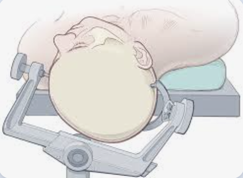
The patient is positioned supine on the table with the shoulder elevated on a roll and the head turned 45° contralateral to the surgical side.
Rey-Dios and Cohen-Gadol apply some degree of head extension to facilitate access to the superior portion of the insula under the frontoparietal operculum. This positioning method allows for a better dissection of the sylvian fissure as it facilitates the opercula to separate/fall awayunder the action of gravity and provides a more accessible trajectory toward the most posterior portion of the insula.
The desired degree of head rotation must be trialed before excessive narcotics and sedation have been administered. If the patient has significant discomfort with the degree of rotation that the surgeon desires, a comfortable wedge of foam can be placed under the torso on the contralateral side to compensate for the lack of neck mobility. In the awake patient, the table is bent into a “beach chair” configuration to optimize patient comfort. Prior to placement of the skull clamp, 0.5% lidocaine with epinephrine and 0.25% bupivacaine in a 1:1 proportion is used to infiltrate the trajectories of the supraorbital and occipital nerves, the incision line, the root of the temporalis muscle, and pin sites. Regional scalp anesthesia will provide additional patient comfort. Whenever available, frameless stereotactic navigation should be used to localize the tumor on the surface to ensure that the scalp flap and craniotomy are large enough to generously expose the tumor and neighboring cortex to be mapped. Once resection has started, the reliability of neuronavigation diminishes due to brain shift, but for the less experienced surgeon, it can remain helpful for orientation during resection. A “trauma flap” or question mark incision is most often used. This incision allows access to the entire length of the sylvian fissure and permits mapping of the language and sensorimotor cortex. The posterior extension of the craniotomy can be tailored based on neuronavigation data. Once the scalp flap is reflected, further infiltration of the temporalis muscle is necessary. Upon elevation of the bone flap, the dura is infiltrated with 0.5% lidocaine with a very fine needle following the trajectory of the middle meningeal artery and radially along the craniotomy edge. If the patient had to be deeply sedated for craniotomy, the process of reawakening should take place after sylvian fissure dissection has been completed and the lateral portion of the insular tumor through the transsylvian approach has been removed 1).
Approaches
Awake surgery for insular glioma
Awake surgery for insular glioma.
Shawn Hervey-Jumper and Berger from the UCSF Medical Center reviewed the literature for published reports focused on insular region anatomy, neurophysiology, surgical approaches, and outcomes for adults with who grade II-IV gliomas.
While originally considered to pose too great a risk, insular glioma surgery can be performed safely due to the collective efforts of many individuals. Similar to resection of gliomas located within other cortical regions, maximal resection of gliomas within the insula offers patients greater survival time and superior seizure control for both newly diagnosed and recurrent tumors in this region. The identification and the preservation of M2 perforating and lateral lenticulostriate artery are critical steps to preventing internal capsule stroke and hemiparesis. The transcortical approach and intraoperative mapping are useful tools to maximize safety.
The insula's proximity to middle cerebral and lenticulostriate arteries, primary motor areas, and perisylvian language areas makes accessing and resecting gliomas in this region challenging. Maximal safe resection of insular gliomas not only is possible but also is associated with excellent outcomes and should be considered for all patients with low- and high-grade gliomas in this area 2).
Advances in microsurgical anatomy and brain mapping techniques have allowed an increase in the extent of resection with acceptable morbidity rates. Transsylvian and transcortical approaches constitute the main surgical corridors, the latter providing considerable advantages and a high degree of reliability. Nevertheless, both surgical corridors yield remarkable difficulties in reaching the most posterior insular region.
Small deep infarcts constitute a well-known risk of motor and speech deficit in insulo-opercular glioma surgery. However, the risk of cognitive deterioration in relation to stroke occurrence in so-called silent areas is poorly known.
In a paper, Loit et al. propose to build a distribution map of small deep infarcts in glioma surgery, and to analyze patients' cognitive outcome in relation to stroke occurrence.
They retrospectively studied a consecutive series of patients operated on for a diffuse glioma between June 2011and June 2017. Patients with lower-grade glioma were cognitively assessed, both before and 4 months after surgery. Areas of decreased apparent diffusion coefficient (ADC) on the immediate postoperative MRI were segmented. All images were registered in the MNI reference by ANTS algorithm, allowing to build a distribution map of the strokes. Stroke occurrence was correlated with the postoperative changes in semantic fluency score in the lower-grade glioma cohort.
One hundred fifteen patients were included. Areas of reduced ADC were observed in 27 out of 54 (50%) patients with a lower-grade glioma, and 25 out of 61 (41%) patients with a glioblastoma. Median volume was 1.6 cc. The distribution map revealed five clusters of deep strokes, corresponding respectively to callosal, prefrontal, insulo-opercular, parietal, and temporal tumor locations. No motor nor speech long-term deficits were caused by these strokes. Cognitive evaluations at 4 months showed that the presence of small infarcts correlated with a slight decrease of semantic fluency scores.
Deep small infarcts are commonly found after glioma surgery, but their actual impact in terms of patients' quality of life remains to be demonstrated. Further studies are needed to better evaluate the cognitive consequences-if any-for each of the described hotspots and to identify risk factors other than the surgery-induced damage of microvessels 3).
Sylvian Fissure Dissection
Sylvian fissure dissection provides a narrow corridor for resection of most insular tumors. In addition, MCA branches often tether the frontal lobe to the temporal lobe, limiting elevation of the frontal lobe and undermining of its operculum to remove tumor underneath the frontoparietal operculum. Since these tumors often have temporal or frontal extensions, additional cortical incisions/resections within the frontal and temporal opercula are necessary to allow the surgeon enough space to maximize tumor excision. Therefore, mapping of the face area (dominant and nondominant tumors) or Broca and Wernicke areas (dominant tumors), is necessary to further guide the location of the safe cortical incision(s) in the inferior frontal and superior temporal gyri to extend the working space and angles to facilitate further tumor exposure 4).
Videos
Awake Brain Mapping in Dominant Side Insular Glioma Surgery: 2-Dimensional Operative Video 5).

