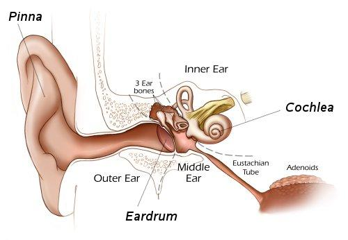
Cochlea
 The cochlea is the auditory portion of the inner ear. It is a spiral-shaped cavity in the bony labyrinth, in humans making 2.5 turns around its axis, the modiolus.
Function
Dedicated to hearing; converting sound pressure patterns from the outer ear into electrochemical impulses which are passed on to the brain via the auditory nerve.
A core component of the cochlea is the Organ of Corti, the sensory organ of hearing, which is distributed along the partition separating fluid chambers in the coiled tapered tube of the cochlea. The name is derived from the Latin word for snail shell, which in turn is from the Greek κοχλίας kokhlias (“snail, screw”), from κόχλος kokhlos (“spiral shell”) in reference to its coiled shape; the cochlea is coiled in mammals with the exception of monotremes.
The cochlear nerve carries auditory sensory information from the cochlea of the inner ear directly to the brain stem. The other portion of the vestibulocochlear nerve is the vestibular nerve, which carries spatial orientation information to the brain from the semicircular canals.
A study was done to examine the relationships of the cochlea as a guide for avoiding both cochlear damage with loss of hearing in middle fossa approaches and injury to adjacent structures in approaches directed through the cochlea.
Twenty adult cadaveric middle fossae were examined using magnifications of ×3 to ×40.
The cochlea sits below the floor of the middle fossa in the area between and below the labyrinthine segment of the facial nerve and greater petrosal nerve (GPN) and adjacent to the lateral genu of the petrous carotid. Approximately one-third of the cochlea extends below the medial edge of the labyrinthine segment of the facial nerve, geniculate ganglion, and proximal part of the GPN. The medial part of the basal and middle turns are the parts at greatest risk in drilling the floor of the middle fossa to expose the nerves in middle fossa approaches to the internal acoustic meatus and in anterior petrosectomy approaches. Resection of the cochlea is used selectively in extending approaches through the mastoid toward the lateral edge of the clivus and front of the brainstem.
An understanding of the location and relationships of the cochlea will reduce the likelihood of cochlear damage with hearing loss in approaches directed through the middle fossa and reduce the incidence of injury to adjacent structures in approaches directed through the cochlea 1).