LINC01783 Promotes Glioma Tumorigenesis by Enhancing GATA3 Expression Through CBP-Mediated H3K27 Acetylation to Suppress PTEN Expression

LINC01783

Type of Study: In vitro and in vivo molecular mechanistic investigation First Author: Shaocai Hao et al. Author Affiliations:

  • Department of Neurosurgery, General Hospital of Ningxia Medical University, Yinchuan, Ningxia, China

Journal: Biofactors DOI: 10.1002/biof.70029 PMID: 40546096 Publication Date: May–June 2025 Title: LINC01783 Promotes Glioma Tumorigenesis by Enhancing GATA3 Expression Through CBP-Mediated H3K27 Acetylation to Suppress PTEN Expression

To elucidate the oncogenic function of the long intergenic non-coding RNA LINC01783 in glioma progression, focusing on its effect on GATA3 expression and PTEN suppression via CBP-mediated H3K27 acetylation.

LINC01783 is significantly upregulated in glioma tissues and enhances glioma progression by promoting GATA3 expression through CBP-mediated H3K27 acetylation. This, in turn, transcriptionally represses PTEN, contributing to increased tumor cell proliferation and stemness.

  • Sample opacity: No clear details on glioma sample number, subtype stratification, or clinical metadata; undermines reproducibility and clinical significance.
  • In vivo data insufficiently controlled: No information on animal randomization, group sizes, or blinding procedures. Xenograft conclusions are weakly supported.
  • Epigenetic mechanistic oversimplification: Attribution of GATA3 regulation solely to CBP-H3K27ac is unconvincing; alternative pathways and compensatory mechanisms are unexamined.
  • Lack of causal proof: The PTEN axis is emphasized, but whether GATA3 mediates all observed phenotypes is not demonstrated.
  • No translational bridge: No therapeutic agent, inhibitor, or antisense strategy explored. The leap to “potential therapeutic target” is scientifically unfounded.
    Read more

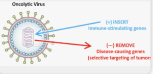
Oncolytic virus‑mediated immunomodulation in glioblastoma: Insights from clinical trials and challenges

In a Review Raziye Piranlioglu *et al.* from

Affiliations Harvey Cushing Neuro‑oncology Laboratories, Dept. Neurosurgery, Brigham and Women’s Hospital, Boston, MA, USA; Dana‑Farber Cancer Institute, Boston, MA, USA

published in *Seminars in Immunology* with the Purpose to synthesize data from clinical trials of oncolytic viruses (OVs) in glioblastoma, evaluating immunomodulatory effects, delivery strategies, and challenges in assessing immune responses. They concluded that Oncolytic virus therapy is well tolerated in GBM trials and can convert the immunosuppressive microenvironment into an immunologically active state. However, limitations in post‑treatment sampling and delivery methods impede full understanding of biological mechanisms.


This review is a rehash of well‑known take‑home messages, offering little in the way of novel synthesis or incisive critique. The authors lean heavily on canonical trials (e.g., oHSV, adenovirus) but fail to integrate preclinical correlates from myeloid-targeting strategies, such as macrophage polarization dynamics or MDSC modulation. There’s no fresh mechanism, no meta‑analysis of response rates, and no exploration of why most trials remain phase I with limited impact. Sample‑scarcity is once again highlighted as a blocker—but no alternative trial designs (e.g., neoadjuvant window cohorts, liquid biopsy) are proposed. In short, the review scratches the surface of challenges without pushing the field forward.

Read more

Maximizing Tumor Resection and Managing Cognitive Attentional Outcomes: Measures of Impact of Awake Surgery in Glioma Treatment

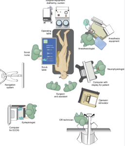
In a retrospective observational study Zigiotto et al. from the S. Chiara University-Hospital, Azienda Provinciale per i Servizi Sanitari, Trento, published in the Neurosurgery Journal on 64 glioma patients who underwent awake surgery (AwS) or asleep surgery (AsS), with neuropsychological and imaging follow-up. They evaluated the impact of awake surgery on attentional outcomes in glioma patients, and analyzed whether greater extent of tumor resection correlates with transient cognitive (attentional) decline, especially in relation to lesions within the default mode network. Awake surgery allows for more extensive supramaximal resection and is associated with longer overall survival, particularly in patients with glioblastomas. However, it also leads to a higher rate of transient postoperative attentional dysfunction, likely due to resection in attention-related brain networks. The study suggests that patient selection and intraoperative cognitive monitoring should be optimized in future glioma surgery 5).


This retrospective study compares awake versus asleep craniotomy in 64 glioma patients, using simple attention tests before and after surgery. The authors claim that awake craniotomy (AwC) allows more extensive tumor resection and leads to longer survival, albeit at the cost of transient attentional dysfunction.

The title promises a nuanced exploration of cognitive outcomes. What it delivers is a reduction of “attention” to the Trail Making Test Part A and a visual search task — an embarrassingly narrow lens for such a multidimensional construct. The study purports to evaluate the impact of surgery on attention, yet fails to define attention, stratify its subtypes, or provide any neuropsychological depth. This is not a cognitive study — it’s a surgical paper pretending to be one.

Read more

Sodium MRI in Pediatric Brain Tumors

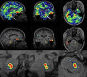
In a narrative review Bhatia et al. from the Children’s Hospital of Philadelphia, Radiological Sciences Laboratory, School of Medicine, Stanford University, published in the American Journal of Neuroradiology to explore the potential of sodium-23 MRI (^23Na-MRI) as a noninvasive imaging modality to assess physiological and biochemical changes in pediatric brain tumors and concluded that is a promising, noninvasive imaging modality capable of providing unique physiological and biochemical information that is not accessible through conventional MRI techniques

This narrative review attempts to position ^23Na-MRI as a frontier imaging technique for pediatric brain tumors. It lauds the modality’s potential to reveal sodium-dependent physiological alterations — but quickly devolves into technological evangelism with minimal clinical anchoring. The piece is high on optimism, low on pragmatism, and entirely devoid of data-supported clinical outcomes.

The article enthusiastically describes the theoretical virtues of sodium MRI — sensitivity to cell integrity, ionic gradients, extracellular space — but offers no compelling clinical cases, no comparative metrics, and no outcome data. What remains is a speculative wish list, presented as a roadmap. The authors confuse imaging potential with diagnostic utility, a common pitfall in radiology reviews driven by physics rather than patient care.

“Exciting” is not a scientific category.

Read more

High-Resolution MR Imaging of the Parasellar Ligaments

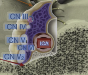
In a anatomic-imaging correlation study with a single-case MR + dissection design Mark el al. 1) from the Department of Radiology, Mayo Clinic, Rochester, Minnesota, USA, Department of Neurosurgery, University of Valencia and Fundación Instituto Valenciano de Oncología (IVO), Valencia, Spain publiseh in the American Journal of Neuroradiology (AJNR) to determine whether high-resolution T2-weighted MRI can visualize the parasellar ligaments, which have previously only been described in cadaveric dissection or intraoperative findings, and to correlate these MRI findings with anatomical dissection in the same specimen. The authors report that parasellar ligaments can be identified on high-resolution T2-weighted MRI as T2-hypointense, bandlike structures originating from the medial wall of the cavernous sinus. They claim that identifying these ligaments may be relevant, given that resection of the medial wall of the cavernous sinus has been associated with better outcomes in functioning pituitary adenoma surgery.


This study is a prime example of technological overreach dressed up as discovery. It takes a single cadaver, applies ultra-high-resolution MRI, and then retrofits a minor fibrous band into a clinical “finding.” The result is a beautifully imaged, clinically irrelevant piece of anatomical embroidery that contributes nothing actionable to radiologyneurosurgery, or pituitary surgery.

❌ Critical Flaws

Read more

Preoperative Nomogram-Based Assessment to Identify GBM Patients Who Do not Derive Survival Benefit From GTR Compared to STR

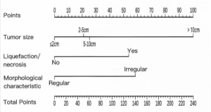
In a retrospective prognostic modeling study, He et al. from Sichuan Provincial People’s Hospital published in the Academic Radiology a preoperative nomogram to identify glioblastoma patients who do not derive a survival benefit from gross total resection (GTR) compared to subtotal resection (STR), and concluded that patients with nomogram scores below 55 or above 95 gain limited survival advantage from GTR, supporting a more individualized surgical strategy 16).


🎯 Takeaway Message for Neurosurgeons

Don’t let a nomogram tell you not to operate. This study reduces complex glioblastoma surgery to a score — ignoring tumor location, function, biology, and patient context. Use it, at best, as background noise. Surgical judgment, not predictive modeling, should guide the extent of resection. Maximal safe resection remains the standard — not because of scores, but because leaving tumor behind costs lives.

Read more

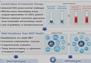
Comparison of survival benefit and safety profile between lenvatinib and donafenib as conversion therapy in patients with hepatocellular carcinoma 

In a retrospective comparative cohort study, Hou et al. 1) published in the American Journal of Translational Research, the authors—affiliated with the Department of Oncology, Department of Gynaecology and Obstetrics, and Department of Neurosurgery at Shijiazhuang People’s Hospital (Hebei, China), as well as Beijing Water Conservancy Hospital—compared the survival benefit and safety profile of lenvatinib versus donafenib as conversion therapy in patients with hepatocellular carcinoma (HCC) at China National Liver Cancer (CNLC) stages I–III.


Lenvatinib demonstrated significantly superior survival outcomes—both in overall survival and progression-free survival—compared to donafenib. It also showed better tolerability, with fewer grade ≥3 adverse events.


❌ 1. Study Design: Retrospective = Weak Evidence

This is yet another retrospective single-center analysis, plagued by inherent biases—selection, reporting, and confounding—that no amount of statistical massaging can resolve. No randomization, no blinding, and no control for treatment timing or physician discretion. In oncology, where treatment nuances matter, such designs should be considered hypothesis-generating at best, not guidance for clinical practice.

Read more

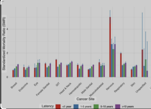
Temporal trends and risk factors associated with stroke mortality among cancer patients

In a retrospective cohort study published in the Journal of Clinical Neuroscience, Ahmed et al. with Cleveland Clinic Cerebrovascular Center, West Virginia University participation 1) analyzed data from over 5.9 million patients diagnosed with a first primary cancer, based on the SEER database (2000–2020). The study aimed to quantify the risk of stroke-related death (SD) in cancer patients and to identify temporal trends and associated clinical and demographic risk factors. Stroke-related mortality (SD) among cancer patients has significantly declined over the past two decades across all cancer types and both sexes. However, older age, non-white race, male sex, and specific cancer types—notably nervous system, respiratory, and head and neck cancers—are associated with a higher risk of stroke death. Conversely, patients receiving chemotherapy or radiotherapy had a lower risk of SD compared to those who received no treatment.


⚠️ Fatal Methodological Flaws

No Clinical Stroke Classification

The authors report on “stroke mortality” without differentiating ischemic vs. hemorrhagic strokes, nor providing stroke etiology or timing relative to cancer diagnosis or cancer treatment—rendering any mechanistic or preventative inference purely speculative.

Read more

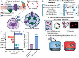
Engineering of CD63 Enables Selective Extracellular Vesicle Cargo Loading and Enhanced Payload Delivery.

In a preclinical experimental study, Obuchi et al. (2025)—with contributions from the Department of Neurosurgery at Leiden University Medical Center—engineered extracellular vesicles for selective cargo loading and enhanced functional delivery, using a modified CD63 scaffold and VSV-G fusion, with in vivo validation in mouse brain models. 1)


🚫 1. Rebranding Complexity as Innovation The authors tout a modular EV engineering system using CD63, mCherry, FLAG-tags, nanobody fusions, and VSV-G. But this is not scientific ingenuity—it’s molecular bricolage. Each component is repurposed from older literature and glued together without real conceptual novelty. The result? A bloated acronym soup with more moving parts than scientific value.

⚠️ What’s pitched as a breakthrough is closer to a tech demo in search of a clinical rationale.

❌ 2. Absence of Disease-Relevant Application Despite name-dropping CRISPR, Cre, and Cas9, no disease context is addressed. No glioma model. No neurodegenerative target. No proof that the cargo accomplishes anything biologically meaningful in the recipient tissue. The mouse brain “validation” is just a fluorescent readout, not a therapeutic outcome.

The cargo arrives, but so what? This is payload delivery without a payload purpose.

🧪 3. Methodological Blind Spots No quantification of EV heterogeneity or functional subpopulations.

No rigorous comparison with alternative delivery systems (e.g., AAVs, lipid nanoparticles).

No evidence of endosomal escape for actual cytoplasmic/nuclear action.

No dose-response curves, toxicity profiling, or repeatability metrics.

This is a biotech prototype, not a therapy-in-the-making.

🔥 4. VSV-G: The Short-Term High, Long-Term Problem The use of VSV-G, a viral fusogen with broad tropism and high immunogenicity, is particularly careless. While it boosts in vitro uptake and helps “sell” delivery efficiency, it introduces a critical translational liability: poor specificity, potential immune activation, and unsuitability for clinical use.

❝Putting VSV-G on EVs is like installing a rocket engine on a paper boat—it moves faster, but it’s doomed to burn out or sink.❞

🧱 5. Structural Inefficiency and Complexity The system requires:

Engineering CD63 with dual tags

Fusing cargo to a nanobody

FLAG-based purification

VSV-G pseudotyping

TEV protease cleavage

This is logistically unscalable for clinical or industrial production and riddled with points of failure. The more components you bolt on, the more it resembles a lab curiosity, not a deliverable platform.

📉 6. Journal Inflation and Institutional Complacency The publication in J Extracell Vesicles is not a mark of impact, but rather a reflection of how EV journals have drifted into translational cosplay, applauding synthetic elegance over clinical relevance. The heavyweight affiliations (MGH, Harvard, etc.) likely ensured acceptance despite the absence of therapeutic depth or mechanistic rigor.

🧠 Conclusion: A study more interested in showing what’s technically possible than what’s biologically meaningful. It trades therapeutic relevance for engineering flair, while ignoring the hard questions of targeting, safetyscalability, and necessity.

This is not a step toward Evidence-based medicine—it’s a flashy side road to nowhere.


1)

Obuchi W, Zargani-Piccardi A, Leandro K, Rufino-Ramos D, Di Lanni E, Frederick DM, Maalouf K, Nieland L, Xiao T, Repiton P, Vaine CA, Kleinstiver BP, Bragg DC, Lee H, Miller MA, Breakefield XO, Breyne K. Engineering of CD63 Enables Selective Extracellular Vesicle Cargo Loading and Enhanced Payload Delivery. J Extracell Vesicles. 2025 Jun;14(6):e70094. doi: 10.1002/jev2.70094. PMID: 40527733.

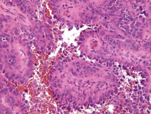
Epithelioid angiosarcoma of the cervical spine: A case report.

Nan et al. 1) describe a rare case of epithelioid angiosarcoma (EA) involving the cervical spine, presenting with pathological fracture and kyphotic deformity, and document the surgical and adjuvant management as well as the clinical outcome in the World Journal of Clinical Cases.

1. Predictable Yet Pointless

The authors claim novelty by describing a rare anatomical presentation of EA. However, this degenerates into a predictable narrative with no new pathophysiological insights, no hypothesis generated, and no clinical paradigm challenged. It is the kind of “rare case” that proliferates in low-barrier journals precisely *because* it demands no intellectual risk.

2. Zero Diagnostic Value

The authors bypass the opportunity to deepen our understanding of the radiological-morphological signature of EA in the spine. No comparative imaging, no differential diagnostic flowchart, no histopathological discussion beyond standard CD31/CD34 immunostaining. If this case had been published in 1995, it would be equally uninformative.

3. Therapeutic Confusion Disguised as Aggressiveness

Two major spine surgeries (posterior decompression + anterior corpectomy) followed by immediate radiotherapy in a moribund patient demonstrate therapeutic overreach without oncological strategy. There is no discussion on multidisciplinary planning, palliative thresholds, or whether delaying surgery or avoiding the second procedure might have prevented ARDS. The reader is left with the impression of a surgical reflex, not an evidence-based decision.

4. No Discussion of Differential Diagnosis or Biomarkers

In a tumor type notorious for being misdiagnosed as metastasischordoma, or sarcoma NOS, the absence of a differential diagnostic framework or advanced markers (ERG, FLI1, HHV-8, etc.) is alarming. Histological laziness cloaked in “rare disease” rhetoric.

5. Outcome Reporting: Conveniently Truncated

The patient dies 3 weeks after surgery, yet the discussion fails to draw any causal or cautionary link between the interventions and the fatal ARDS. No autopsy data, no postmortem imaging, no pulmonary workup. This omission sterilizes the clinical narrative, reducing it to anecdote.

6. Ethically Murky

The case implicitly raises an ethical dilemma—should maximal surgery be performed in aggressive, terminal tumors without demonstrated systemic control? Yet the authors shy away from even mentioning this, let alone framing it for academic discussion.

7. Journal Choice Reflects the Paper’s Weakness

Published in a journal known for minimal peer review stringency, the article offers no citations of recent molecular or targeted therapy advances, no engagement with broader oncological guidelines, and no rationale for the treatment decisions beyond procedural listing.

  • Histological laziness: Failing to provide in-depth pathology discussion beyond CD31/CD34 and H&E staining in vascular tumors.
  • Surgical reflex: The tendency to operate based on mechanical findings (compression, fracture) without integrating prognosis or systemic disease behavior.
  • Ethical sterilization: Avoiding uncomfortable questions about futility, risk-benefit tradeoffs, and overtreatment in end-stage patients.
  • Postmortem evasion: Reporting a perioperative death without diagnostic closure (autopsy, imaging, or medical reflection).

This case report is an example of procedural reporting devoid of scientific merit, clinical reflection, or ethical introspection. It contributes nothing to the understanding of EA, its diagnosis, biology, or management—beyond reiterating its rarity. In its current form, it is neither hypothesis-generating nor practice-changing, and serves as a cautionary tale on how not to write a case report.

  • Include comparative radiology with metastatic disease and primary bone tumors.
  • Provide autopsy findings or detailed explanation of respiratory decline.
  • Discuss therapeutic alternatives (e.g., single-stage surgery, biopsy + RT, palliative care).
  • Frame the case within an oncological decision-making algorithm.

1)

Nan YH, Chiu CD, Chen WL, Chen LC, Chen CC, Cho DY, Guo JH. Epithelioid angiosarcoma of the cervical spine: A case report. World J Clin Cases. 2025 Jun 16;13(17):101593. doi: 10.12998/wjcc.v13.i17.101593. PMID: 40524767; PMCID: PMC11866273.