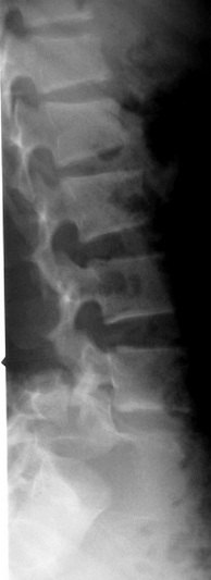
Traumatic Spondyloptosis
Spondyloptosis is the most severe translation spine injury. It results in complete disruption of the structural elements of the vertebral column and the adjacent paravertebral soft tissues, culminating in severe biomechanical instability.
In this injury, all 3 spinal columns are disrupted and show complete loss of alignment.
Etiology
Spondyloptosis usually results from high-velocity injury, and many patients will also have other associated injuries.
Cervical spondyloptosis
Spondyloptosis of the cervical spine at C6-7 level with cord compression is described in a 51-year-old male. Because the bodies of C6 and 7 were tightly locked together, cervical traction failed. Then the patient was operated on by a posterior approach. Posterior stabilization and fusion were performed by C4-5 lateral mass and C7-T1 pedicle screw fixation and rod instrumentation with bridging both C4-5's rods to the C7-T1's extended ones. After C6 total laminectomy and foraminotomy, the C6 body was returned to its proper position. Secondly, anterior stabilization and fusion were performed by C6-7 discectomy with a screw-plate system. A postoperative lateral plain radiograph showed good realignment. In this case, we report the clinical presentation and discuss the surgical modalities of C6-7 total spondyloptosis and the failed close reduction 1).
Thoracic spondyloptosis
Lumbosacral Spondyloptosis
Case series
In total, 20 patients with spondyloptosis were treated during the period reviewed. The mean age of the patients was 27 years (range 12-45 years), and 17 patients were male (2 boys and 15 men) and 3 were women. Fall from height (45%) and road traffic accidents (35%) were the most common causes of the spinal injuries. The grading of the American Spinal Injury Association (ASIA) was used to assess the severity of spinal cord injury, which for all patients was ASIA Grade A at the time of admission. In 11 patients (55%), the thoracolumbar junction (T10-L2) was involved in the injury, followed by the dorsal region (T1-9) in 7 patients (35%); 1 patient (5%) had lumbar and 1 patient (5%) sacral spondyloptosis. In 19 patients (95%), spondyloptosis was treated surgically, involving the posterior route in all cases. In 7 patients (37%), corpectomy was performed. None of the patients showed improvement in neurological deficits. The mean follow-up length was 37.5 months (range 3-60 months), and 5 patients died in the follow-up period from complications due to formation of bedsores (decubitus ulcers).
To the authors' best knowledge, this study was the largest of its kind on traumatic spondyloptosis. Its results illustrate the challenges of treating patients with this condition. Despite deformity correction of the spine and early mobilization of patients, traumatic spondyloptosis led to high morbidity and mortality rates because the patients lacked access to rehabilitation facilities postoperatively 2).
Case reports
A 28-year-old man presented with severe low back pain, numbness at the soles of feet, and bowel and bladder dysfunction. Two days before admission, a tree trunk fell on his back while he was seated. A two-stage posterior-anterior procedure was performed. At the first stage, posterior decompression, reduction, and fusion with instrumentation were performed. At the second stage, which was performed 6 days after the first stage, the patient underwent anterior lumbar interbody fusion. The patient received physical therapy 1 week after the second stage. Results The patient's numbness improved immediately after the first posterior surgery. His fecal and urinary incontinence improved 6 months after discharge. He has been pain-free for a year and has returned to work.
A PubMed search was performed using the following keywords: lumbosacral spondyloptosis, lumbosacral dislocation, and L5-S1 traumatic dislocation. The search returned only nine reported cases of traumatic spondyloptosis. Traumatic spondyloptosis at the lumbosacral junction is a rare ailment that should be suspected in cases of high, direct, and posterior impact on the low lumbar area, and surgical treatment should be the standard choice of care 3).
