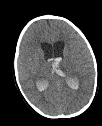
Traumatic intraventricular hemorrhage
Most studies of traumatic intraventricular hemorrhage (tIVH) contain few subjects and are retrospective in design, providing minimal information about the entity and its clinical significance.
Epidemiology
Most of these cases were caused by traffic accident 1).
The incidence of IVH in nonpenetrating head injury is 1.5 to 3% and 10 to 25% of patients with severe head injury (GCS less than 8) have IVH 2) 3) 4) 5).
Etiology
The actual origin of IVH has rarely been identified and the pathogenesis and mechanisms involved are still obscure.
Shear strain in severe head injury should cause tears of the subependymal vein, fornix, septum pellucidum and choroid plexus. 8 of Hashimoto et al. cases out of 32 ventricular hemorrhage showed tear of the subependymal vein. 8 of the cases showed damage of the fornix of septum pellucidum. These results suggested that the anatomical structure of the fornix and septum pellucidum were weak points for shearing force. 20 cases in 32 with ventricular hemorrhage died. These cases were frequently associated with other traumatic lesions, namely contusion of white matter and grey matter, brain stem lesions and cerebellar contusion, caused by shearing injury. Therefore, the prognosis of severe head injury with intraventricular hemorrhage is poor 6).
Types
Hashimoto et al., divided traumatic intraventricular hemorrhage into 4 types.
Type 1: massive hemorrhage (9 cases).
Type 2: subependymal hemorrhage (8 cases).
Type 3: damage of fornix or septum pellucidum (8 cases).
Type 4: Nieveau (7 cases). 7).
Outcome
Poor prognosis in this condition is a reflection of the severity of the injury to the head. Intracerebral hemor- rhage (ICH) and basal ganglia hemorrhage usually find a way to the ventricles.
In the absence of intraparenchymal hemorrhage IVH is most often caused by tearing of the subependymal veins in the fornix, septum pellucidum or choroid plexus found in autopsy studies.
Posttraumatic IVH rarely produces hydrocephalus, and the presence of IVH in a patient with image-proven diffuse axonal injury (DAI) shows that other pathological mechanisms are involved.
Prognosis is poor in these patients and it is not known whether it is due to the presence of blood in the ventricle per se, due to induced hydrocephalus or increased intracranial pressure.
Case series
2006
Atzema et al. prospectively enrolled trauma patients from 18 centers in North America in the National Emergency X-Radiography Utilization Study (NEXUS) II if they received an emergent head computed tomography (CT) scan, as determined by the managing physician. Clinical data were collected at the time of enrollment and CT reports were compiled at least 1 month later. They calculated prevalence and demographics of tIVH from the 18 sites, while outcome data were gathered from medical records of patients with tIVH who were seen at any of six sites that participated in the follow-up portion of the study. They considered patients who underwent a neurosurgical intervention or who had a “poor outcome” (Glasgow Outcome Scale score of 1 to 3, death, persistent vegetative state, or severe disability) to have suffered a “combined outcome.”
Prevalence of tIVH among all trauma patients who received a head CT was 118 in 8,374, or 1.41%. Among tIVH patients, 70% had a “poor outcome” and 76% had a “combined outcome.” A poor outcome appeared to be associated with an abnormal presenting Glasgow Coma Scale score and involvement of the third or fourth ventricle, whereas age appeared to be unrelated. Patients with tIVH and no major associated injury on CT tended to do well; only one patient with isolated tIVH had a poor outcome.
Traumatic IVH is rare and is associated with poor outcomes that seem to be the consequence of associated injuries. Isolated tIVH patients who are clinically well appear to have a functional outcome; we were unable to identify a case of isolated tIVH, combined with a normal neurologic examination, resulting in a poor or combined outcome 8).
1992
Among 329 cases with comatose state caused by severe head injury, 32 had primary intraventricular hemorrhage as revealed on initial CT scan (13.4%).
Hashimoto et al., divided traumatic intraventricular hemorrhage into 4 types. Type 1: massive hemorrhage (9 cases). Type 2: subependymal hemorrhage (8 cases). Type 3: damage of fornix or septum pellucidum (8 cases). Type 4: Nieveau (7 cases). Most of these cases were caused by traffic accident. Shearing injury may be the most accurate mechanism to produce the intraventricular hemorrhage. Shear strain in severe head injury should cause tears of the subependymal vein, fornix, septum pellucidum and choroid plexus. 8 cases out of 32 ventricular hemorrhage showed tear of the subependymal vein. 8 of our cases showed damage of the fornix of septum pellucidum. These results suggested that the anatomical structure of the fornix and septum pellucidum were weak points for shearing force. 20 cases in 32 with ventricular hemorrhage died. These cases were frequently associated with other traumatic lesions, namely contusion of white matter and grey matter, brain stem lesions and cerebellar contusion, caused by shearing injury. Therefore, the prognosis of severe head injury with intraventricular hemorrhage is poor 9).
Before the advent of computed tomography, intraventricular hemorrhage (IVH) from any source was thought rare and invariably fatal. Although intraventricular blood is readily identifiable with computed tomography, there has been little systematic study of its significance in blunt head trauma. Forty-three patients with traumatic IVH were prospectively identified in 1 year at Harborview Medical Center (University of Washington). Most were victims of motor vehicle accidents and suffered severe head injuries. IVH occurred alone in two patients; superficial contusions and subarachnoid hemorrhage were the most common associated finding. Blood was present in only one or both lateral ventricles in 25 patients; only the 3rd or 4th ventricles in 4 and all ventricles in 14 instances. There were 3 intracerebral hematomas and 14 basal ganglion hemorrhages. All of the former and half of the latter communicated with the adjacent lateral ventricle. Extra-axial hematomas appeared more common when only the lateral ventricles were involved, whereas corpus callosum or brain-stem hemorrhage appeared more likely when all the ventricles were involved. Acute hydrocephalus was rare, and ventricular drainage was needed in only four cases. Intracranial pressure (ICP) was elevated (> 15 mm Hg) in 46% of patients. The amount of IVH was related inversely with the Glasgow Coma Scale, but not with increased ICP. The presence of IVH indicated a poor outcome, with only half of the patients being independent at a 6-month follow-up. Poor outcome was associated with increasing age, low admission Glasgow Coma Scale, the presence of space occupying lesions if only the lateral ventricles were involved, and hemorrhage in all four ventricles 10).
Case reports
A case of primary post-traumatic intraventricular hemorrhage is presented. The patient, a 22 years-old man, after a mild head injury and a lucid interval lost consciousness and progressively developed symptoms of raised intracranial pressure. CT scanning, 24 hours later, revealed blood in the ventricles without concomitant brain contusion. The patient's condition improved dramatically after an external ventricular drainage 11).
