Thoracic pedicle screw
The use of thoracic pedicle screw instrumentation has become increasingly widespread in the treatment of scoliosis owing to the consistently superior results achieved in terms of fixation and deformity correction 1) 2).
The accuracy of pedicle screws is typically defined as the screws axis being fully contained within the cortices of the pedicle 3) 4).
Placement
Exposure
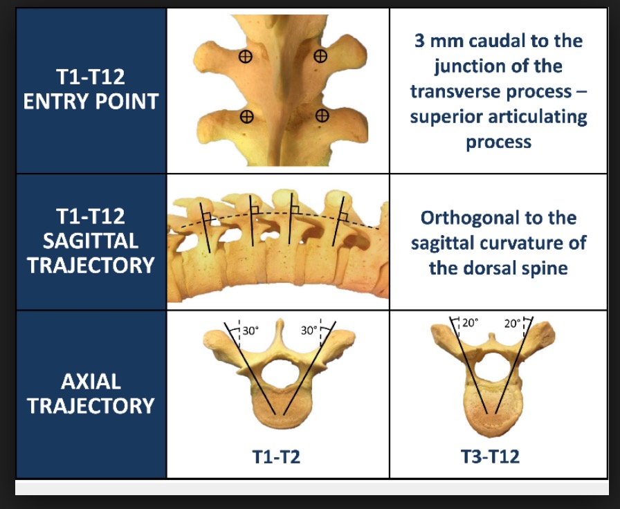
A midline skin incision is made one spinous process above the most cranial thoracic vertebra down to the spinous process of the most caudal vertebra. The extensor muscles are dissected laterally to the tips of the transverse processes. The inferior facet is resected except in the most cranial vertebra, where the capsule is excised only. The lateral border of the superior facet is identified with a probe. A parallel line of the lateral border of the superior facet corresponds to the y-axis of the entry point to the pedicle. The transverse process is divided in three horizontal parts. A parallel line between the middle and the cranial third of the transverse process corresponds to the x-axis. The intersection of both axes does not correspond to the center of the pedicle but to the lateral border of the oval
Entry points
The base of the superior articular process at the junction of the lateral one-third and medial two-thirds can be used as an ideal pedicle entry point
5).
Complications
Pedicle screw instrumentation has become increasingly popular during the past 20 years and a vast selection of products is available on the market. With rising implantation rates, reports about specific complications also have increased. The main reason for these complications is the fact that the course of the pedicle and in turn the positioning of the pedicle screw cannot be adequately controlled visually. Based on the anatomy of the surrounding structures, complications caused by malpositioning can be divided into three main groups: mechanical, neurological and vascular. Beyond mechanical limitations of spinal motion, nerve injury can lead to neurological problems while injuries to vascular structures usually cause hemorrhage. These typical problems in general become apparent intraoperatively or in the immediate postoperative course 6).
see Thoracic screw misplacement.
The term acceptable has been hypothesized mainly in thoracic spine due to the frequent and clinically benign nature of pedicle wall compromise in smaller thoracic pedicles 7).
Malpositioning
Malposition is the most commonly reported complication of thoracic pedicle screw placement, at a rate of 15.7% per screw inserted with postoperative computed tomography scans. The use of pedicle screws in the thoracic spine for the treatment of pediatric deformity has been reported to be safe despite the high rate of patients with malpositioned screws (11%). Major complications, such as neurologic or vascular injury, were almost never reported in this literature review of case series. Cases reports on the other hand have started to identify such complications 8).
Although the clinical course in malpositioned pedicle screw instrumentation may stay unremarkable, in a proven injury to the thoracic aorta revision is mandatory to prevent further vascular damage. The appropriate strategy demands exact and provident planning using a preferably interdisciplinary approach 9).
Reports of the accuracy of existing neuromonitoring methods for detecting or preventing medial malpositioning of thoracic pedicle screws have varied widely in their claimed effectiveness.
In a prospective, blinded and randomized study using a novel combination of input (4-pulse stimulus trains delivered within the pedicle track) and output (evoked electromyography from leg muscles) to detect pedicle track trajectories that-once implanted with a screw-would cause that screw to breach the pedicle's medial wall and encroach upon the spinal canal. For comparison, the authors also used screw stimulation as an input and evoked electromyogram from intercostal and abdominal muscles as output measures. Intraoperative electrophysiological findings were compared with postoperative CT scans by multiple reviewers blinded to patient identity or intraoperative findings.
Data were collected from 71 patients, in whom 802 screws were implanted between the T-1 and L-1 vertebral levels. A total of 32 screws ended up with screw threads encroaching on the spinal canal by at least 2 mm. Pulse-train stimulation within the pedicle track using a ball-tipped probe and electromyography from lower limb muscles correctly predicted all 32 (100%) of these medially malpositioned screws. The combination of pedicle track stimulation and electromyogram response from leg muscles proved to be far more effective in predicting these medially malpositioned screws than was direct screw stimulation and any of the target muscles (intercostal, abdominal, or lower limb muscles) we monitored. Based on receiver operating characteristic analysis, the combination of 10-mA (lower alarm) and 15-mA stimulation intensities proved most effective for detection of pedicle tracks that ultimately gave rise to medially malpositioned screws. Additional results pertaining to the impact of feedback of these test results on surgical decision making are provided in the companion report. Conclusions This novel neuromonitoring approach accurately predicts medially malpositioned thoracic screws. The approach could be readily implemented within any surgical program that is already using contemporary neuromonitoring methods that include transcranial stimulation for monitoring motor evoked potentials 10).
Neuromonitoring strategy during placement of thoracic pedicle screws can significantly reduce the incidence of clinically relevant thoracic pedicle screw medial malpositioning 11).
Avoidance
Stereotactic image guidance
Despite the use of adjunctive techniques, accurate pedicle screw placement in thoracic spine remains to be challenging. New technology, such as stereotactic image guidance applied to screw placement has been associated with increased accuracy 12).
However, this technique requires CT scanning with preoperative irradiation and expensive equipment. The free hand pedicle screw insertion technique exhibits similar accuracy in experienced hands as compared to the image-guided techniques 13).
CT-based study demonstrated that T4-T9 concave segments have a smaller safe zone with respect to both cord-aorta injury in medial and lateral malpositions. In these segments, screws should be accurate and screw malposition is to be unacceptable 14).
Biplane fluoroscopy guided robot system (BFRS)
The BFRS might be helpful in improving the accuracy of percutaneous pedicular screw insertion procedures. In the future, we will attempt to improve the accuracy and reliability of the BFRS and to determine new clinical applications for the BFRS 15).
Triggered electromyography (t-EMG)
Independent of the screw position, average t-EMG thresholds were always higher at the convexity (CV) of the scoliotic curves in the apex and above the apex regions, presuming that the distance from the pedicle to the spinal cord plays an important role in electrical transmission. The t-EMG technique has low sensitivity to predict screw malpositioning and cannot discriminate between medial cortex breakages and complete invasion of the spinal canal 16).
When compared with screws made of stainless steel (SS), most titanium alloy (Ti-alloy) pedicle screws behaved more like semiconductors, showing conduction properties that were highly frequency dependent. These properties likely contributed to the difficulties encountered in interpreting thoracic screw placements based on stimulus-evoked electromyography from direct screw stimulation 17).
slide technique
This technique is very close to the “funnel technique”. The “funnel” and then the “slide” technique are mostly useful in complex spinal deformities as in neuromuscular patients. The “slide technique” is a safe, effective and cost-effective technique for pedicle screw placement in the thoracic spine especially in severe deformities. LEVEL OF EVIDENCE: IV 18).

