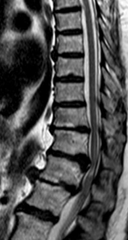
Thoracic disc herniation magnetic resonance imaging
Magnetic Resonance Imaging (MRI) is the most useful imaging tool to identify disc pathology.
MRI: noncontrast thoracic MRI is the mainstay of diagnosis. If MRI is contraindicated, a thoracic CT/myelogram is the second choice.
T2
A 42-year-old male presents with a progressive clinical history of weakness and a sensation of paresthesias in the lower limbs. He describes a pressure sensation in the abdominal region at the T5-T7 level. Over the past 3 days, he has experienced multiple episodes of urinary incontinence and one episode of fecal incontinence. Additionally, his partner reports erectile dysfunction. He came to the Emergency Room due to a new fall caused by instability while dressing.
Broad-based posteromedial disc herniation of the C7/Th1 disc with a component of posteromedial disc extrusion migrated cranially as caudally and with mild indentation on the spinal cord.
Right posterolateral herniation of the Th2/D3 disc with marked indentation on the right anterior aspect of the spinal cord and significant signs of severe vertebral canal stenosis due to facet joint convergence and hypertrophy of yellow ligaments, with altered signal intensity of the spinal cord in relation to established compressive myelopathy.
Broad-based posteromedial protrusion of the D3/D4 disc with moderate involvement of the intervertebral foramina.
Broad-based posteromedial protrusion of the D4/D5 disc, mild.
Left paramedial protrusion of the D7/D8 disc and posteromedial protrusion of the D8/D9 disc indenting on the thecal sac.
Longitudinal dorsal high skin incision exposing C7-Th3. Bilateral skeletonization of Th2-Th3. Canulated screws (S4 Braun Aesculap) 30*4.5 in Th2-Th3 with MNIO control. Posterior decompression due to ligamentous hypertrophy, facet joint issues, and chronic spinal cord compression. Loss of somatosensory-evoked potentials (PESS) and motor-evoked potentials in the middle extremities (MID) during decompression (see report). Continued decompression with motor recovery, performing undercutting of Th1 and Th3 laminae until complete canal liberation. Loss of motor potentials prior to hemostasis (see report).
Hemostasis aided by Floseal. Espongostan on dura. Connection of the system with 35 and 40 bars and autologous bone + demineralized bone matrix (DBM). Fascia and subcutaneous tissue closed with Vicryl. Skin closure with sutures.

