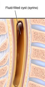
Syrinx fluid
The origin of syrinx fluid is controversial.
Most hypothesis trying to explain the pathophysiology of idiopathic syringomyelia involve mechanisms whereby CSF is pumped against a pressure gradient, from the subarachnoid space into the cord parenchyma. On review, these theories have universally failed to explain the disease process. A few papers have suggested that the syrinx fluid may originate from the cord capillary bed itself. However, in these papers, the fluid is said to accumulate due to impaired fluid drainage out of the cord. Again, there is little evidence to substantiate this. This proffered hypothesis looks at the problem from the perspective that syringomyelia and normal pressure hydrocephalus are almost identical in their manifestations but only differ in their site of effect within the neuraxis. It is suggested that the primary trigger for syringomyelia is a reduction in the compliance of the veins draining the spinal cord. This reduces the efficiency of the pulse wave dampening, occurring within the cord parenchyma, increasing arteriolar and capillary pulse pressure. The increased capillary pulse pressure opens the blood-spinal cord barrier due to a direct effect upon the wall integrity and interstitial fluid accumulates due to an increased secretion rate. An increase in arteriolar pulse pressure increases the kinetic energy within the cord parenchyma and this disrupts the cytoarchitecture allowing the fluid to accumulate into small cystic regions in the cord. With time the cystic regions coalesce to form one large cavity which continues to increase in size due to the ongoing interstitial fluid secretion and the hyperdynamic cord vasculature 1).
Heiss et al., prospectively studied patients with syringomyelia: 22 with Chiari I malformation and 16 with spinal subarachnoid space (SAS) obstruction-related syringomyelia before and 1 wk after surgery, and 9 with tumor-related syringomyelia before surgery only. Computed tomography-myelography quantified dye transport into the syrinx before and 0.5, 2, 4, 6, 8, 10, and 22 h after contrast injection by measuring contrast density in Hounsfield units (HU).
Before surgery, more contrast passed into the syrinx in Chiari I malformation-related syringomyelia and spinal obstruction-related syringomyelia than in tumor-related syringomyelia, as measured by (1) maximum syrinx HU, (2) area under the syrinx concentration-time curve (HU AUC), (3) ratio of syrinx HU to subarachnoid cerebrospinal fluid (CSF; SAS) HU, and (4) AUC syrinx/AUC SAS. More contrast (AUC) accumulated in the syrinx and subarachnoid space before than after surgery.
Transparenchymal bulk flow of CSF from the subarachnoid space to syrinx occurs in Chiari I malformation-related syringomyelia and spinal obstruction-related syringomyelia. Before surgery, more subarachnoid contrast entered syringes associated with CSF pathway obstruction than with tumor, consistent with syrinx fluid originating from the subarachnoid space in Chiari I malformation and spinal obstruction-related syringomyelia and not from the subarachnoid space in tumor-related syringomyelia. Decompressive surgery opened subarachnoid CSF pathways and reduced contrast entry into syringes associated with CSF pathway obstruction 2).
