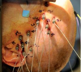
Stereoelectroencephalography electrode implantation
Indications
Among other general indications for invasive intracranial electroencephalography (EEG) monitoring, its advantages include access to deep cortical structures, its ability to localize the epileptogenic zone when subdural grid electrodes have failed to do so, and its utility in the context of possible multifocal seizure onsets with the need for bihemispheric explorations.
Stereoelectroencephalography (sEEG) depth electrodes enable exploration of deeply located structures and lesions, and of buried cortex, which are not easily assessable by subdural or other iEEG methods. Simultaneous recording of SEEG signals from deep and superficial brain structures allows, when the position of each electrode is precisely determined, delineation of a three-dimensional, spatial and temporal organization of epileptic activities 1).
The main advantage is the possibility to study the epileptogenic neuronal network in its dynamic and 3-dimensional aspect, with optimal time and space correlation, with the clinical semiology of the patient's seizures 2).
Stereoelectroencephalography electrode implantation accuracy
see Stereoelectroencephalography electrode implantation accuracy.
Magnetoencephalography and stereoelectroencephalography are often necessary in the course of the non-invasive and invasive presurgical evaluation of challenging patients with drug resistant epilepsy.
In a study, Murakami et al., aim to examine the significance of magnetoencephalography dipole clusters and their relationship to stereo-electroencephalography findings, area of surgical resection, and seizure outcome. They also aim to define the positive and negative predictors based on magnetoencephalography dipole cluster characteristics pertaining to seizure-freedom. Included in this retrospective study were a consecutive series of 50 patients who underwent magnetoencephalography and stereo-electroencephalography at the Cleveland Clinic Epilepsy Center. Interictal magnetoencephalography localization was performed using a single equivalent current dipole model. Magnetoencephalography dipole clusters were classified based on tightness and orientation criteria. Magnetoencephalography dipole clusters, stereo-electroencephalography findings and area of resection were reconstructed and examined in the same space using the patient's own magnetic resonance imaging scan. Seizure outcomes at 1 year post-operative were dichotomized into seizure-free or not seizure-free. We found that patients in whom the magnetoencephalography clusters were completely resected had a much higher chance of seizure-freedom compared to the partial and no resection groups (P = 0.007). Furthermore, patients had a significantly higher chance of being seizure-free when stereo-electroencephalography completely sampled the area identified by magnetoencephalography as compared to those with incomplete or no sampling of magnetoencephalography results (P = 0.012). Partial concordance between magnetoencephalography and interictal or ictal stereo-electroencephalography was associated with a much lower chance of seizure freedom as compared to the concordant group (P = 0.0075). Patients with one single tight cluster on magnetoencephalography were more likely to become seizure-free compared to patients with a tight cluster plus scatter (P = 0.0049) or patients with loose clusters (P = 0.018). Patients whose magnetoencephalography clusters had a stable orientation perpendicular to the nearest major sulcus had a better chance of seizure-freedom as compared to other orientations (P = 0.042). Our data demonstrate that stereo-electroencephalography exploration and subsequent resection are more likely to succeed, when guided by positive magnetoencephalography findings. As a corollary, magnetoencephalography clusters should not be ignored when planning the stereo-electroencephalography strategy. Magnetoencephalography tight cluster and stable orientation are positive predictors for a good seizure outcome after resective surgery, whereas the presence of scattered sources diminishes the probability of favourable outcomes. The concordance pattern between magnetoencephalography and stereo-electroencephalography is a strong argument in favour of incorporating localization with non-invasive tools into the process of presurgical evaluation before actual placement of electrodes 3).
