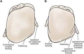
Positional Plagiocephaly Differential Diagnosis
General practitioners (GPs) have an important role in assessment, diagnosis and referral for paediatric skull deformities. GPs are well placed to clinically differentiate between deformational plagiocephaly and craniosynostosis and provide timely referrals to optimise patient outcomes 1).
The child with unilateral lambdoid synostosis has a thick ridge over the fused suture, with compensatory contralateral parietal and frontal bossing 2). There is an ipsilateral occipitomastoid bulge, with a posteroinferior displacement of the ipsilateral ear. These characteristics are opposite to the findings in the children with deformational plagiocephaly. In the view from above, the shape of the head will be trapezoid in lambdoid synostosis and parallelogram in deformational plagiocephaly. A 3D CT will confirm the diagnosis. Torticollis is most commonly associated with deformational plagiocephaly. Chiari malformation can be present with lambdoid synostosis.
