Mastoidectomy
Procedure performed to remove the mastoid air cells. This can be done as part of treatment for mastoiditis, chronic suppurative otitis media or Cholesteatoma. In addition, it is sometimes performed as part of other procedures (cochlear implant) or for access to the middle ear.
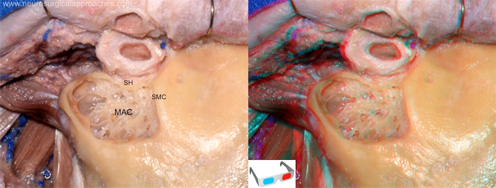
The cortex over the mastoid bone is removed by using a high speed drill with a large cutting bur. The anterior border of the bone removal is a slightly curved line that extends between the top of the external auditory meatus and the mastoid tip. The superior limit is a line perpendicular to the first line, extending from the root of the zygoma posteriorly to the asterion: the supramastoid crest.
The major landmarks in the mastoid area are the lateral semicircular canal, the mastoid antrum superiorly, and the digastric ridge inferiorly. As the cortical bone is drilled, air cells will begin to be encountered.
MAC: mastoid air cells; SH: spine of Henle; SMC: suprameatal crest.
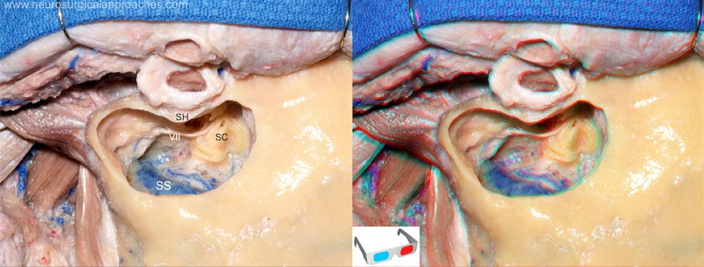
Bone is drilled 1cm behind the sigmoid sinus, maintaining a uniform depth until the sigmoid sinus is exposed. The sigmoid sinus is skeletonized along its length down through the region of the jugular bulb using a diamond burr. The mastoid air cells are removed anteriorly and superiorly to expose the middle fossa dura. The air cells are drilled further anteriorly to expose the compact bone of the bony labyrinth. Once the semicircular canals are exposed we must know that the facial nerve is situated parallel and 1 to 2 mm anterior to the lateral semicircular canal.
SC: Semicircular canals; SH: Spine of Henle; SS: Sigmoid sinus.
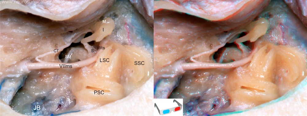
The mastoidectomy has been completed to expose the capsule of the posterior and lateral canals and the tympanic and mastoid facial segments. The mastoid segment of the facial nerve descends through the facial canal and gives rise to the chorda tympani, which passes upward and forward across the tympainc membrane and the malleus neck.
CT: chorda tympani; I: incus; JB: jugular bulb; LSC: lateral semicircular canal; M: malleus; PSC: posterior semicircular canal; S: stapes; SM: stapedius muscle; SSC: superior semicircular canal; VIIms: mastoid segment of VII cranial nerve; VIIts: tympanic segment of VII cranial nerve.
Types
There are classically 5 different types of Mastoidectomy:
Radical Mastoidectomy - Removal of posterior and superior canal wall, meatoplasty and exteriorisation of middle ear. Canal Wall Down Mastoidectomy - Removal of posterior and superior canal wall, meatoplasty. Tympanic membrane left in place. Canal Wall Up Mastoidectomy - Posterior and superior canal wall are kept intact. A facial recess approach is taken. Cortical Mastoidectomy (Also known as schwartze procedure) - Removal of Mastoid air cells is undertaken without affecting the middle ear. This is typically done for mastoiditis Modified Radical Mastoidectomy - This is confusing because it is typically described as a radical mastoidectomy while maintaining the posterior and superior canal wall which reminds the reader of the Canal Wall Up Mastoidectomy. However, the difference is historical. Modified radical mastoidectomy typically refers to Bondy's procedure which involves treating disease affecting only the epitympanum. Diseased areas as well as portions of the adjacent superior and posterior canal are simply exteriorised without affecting the uninvolved middle ear.


