Low-grade glioma old literature
Old definition
Low-grade gliomas (LGGs) are a diverse group of WHO grade I - WHO grade II of glial origin with pilocytic astrocytoma (PA) being the most frequent LGG diagnosis. It is now generally accepted that PA and most other LGGs are a single pathway disease of the MAPK signal transduction pathway 1).
Epidemiology
Low-grade glioma classification
Low-grade astrocytoma
Pilomyxoid astrocytoma 9425/3* - WHO grade II
Pleomorphic xanthoastrocytoma 9424/3 - WHO grade II
Diffuse astrocytoma 9400/3 - WHO grade II
Fibrillary astrocytoma 9420/3
Protoplasmic astrocytoma 9410/3
Gemistocytic astrocytoma 9411/3
e.g. oligoastrocytoma
The term should not be used for a specific, non-infiltrative WHO I tumour of astrocyte-lineage such as pleomorphic xanthoastrocytoma (PXA), subependymal giant cell astrocytoma (SGCA) and pilocytic astrocytoma, as these have different prognosis, treatment and imaging features.
Martino et al. reviewed a consecutive series of 19 patients with GIIG within functional areas who underwent two operations separated by at least 1 year. Intraoperative Electrostimulation mapping was used in all operations for recurrence and in 14 of the initial procedures. A specific rehabilitation was provided. FINDINGS: At the first operation, we performed 14 subtotal and 5 partial resections. Eighteen patients returned to a normal socio-professional life. Nine patients received adjuvant treatment. At the second operation, we performed 1 total, 13 subtotal and 5 partial resections. Three patients with a preoperative neurological deficit improved, 13 remained unchanged, and 3 slight new deficits appeared. In 14 of the 17 patients with preoperative chronic epilepsy, the seizures were reduced or disappeared. Sixteen patients returned to a normal socio-professional life. Pathohistological examination showed that 11 tumours had progressed to high-grade glioma. The median time between the two operations was 4.1 years (range 1 to 7.8 years) and the median follow-up from initial diagnosis was 6.6 years (range 2.3 to 14.3 years). No deaths occurred during the follow-up period. CONCLUSIONS: Repeat operations guided by intra-operative Electrostimulation is an efficacious treatment for recurrent grade II glioma in an eloquent area 2).
Molecular profile
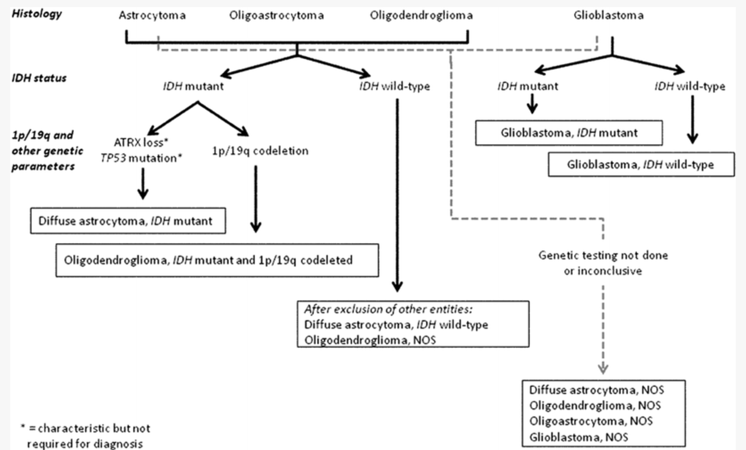
The most critical molecular alterations (IDH1/2, 1p/19q codeletion, ATRX, TERT, CIC, and FUBP1) and circumscribed (BRAF-KIAA1549, BRAF(V600E), and C11orf95-RELA fusion) gliomas. These molecular features reflect tumor heterogeneity and have specific associations with patient outcome that determine appropriate patient management. This has led to an important, fundamental shift toward developing a molecular classification of World Health Organization grade II-III diffuse glioma 3).
Low-grade gliomas (LGG) are classified into three distinct groups based on their IDH1 mutation and 1p/19q codeletion status, each of which is associated with a different clinical expression. The genomic sub-classification of LGG requires tumor sampling via neurosurgical procedures.
With the advance of genomics research, there have been a new breakthrough in the molecular classification of gliomas. Glioblastoma (WHO grade Ⅳ) could be subtyped to proneural, neural, classical, and mesenchymal according to the mRNA expression. Low-grade gliomas (WHO grade Ⅱ and Ⅲ) could be divided into 5 types using 1p/19q co-deletion, isocitrate dehydrogenase(IDH) mutation, and TERTp (promotor region) mutation. In 2016, a new classification of tumors of the central nervous system was proposed, and some new markers such as IDH1 mutation were introduced into the diagnosis of gliomas. Genotype and phenotype were integrated to diagnose gliomas. In the meantime, precision treatment for gliomas has also been vigorously developed 4).
With the increased understanding of glioma tumour genetics there is a need to understand the changes and their implications for patient management. There has also been an increasing trend for adopting earlier, more aggressive surgical approaches to Low-grade glioma treatment 5).
Verma and Mehta et al., discuss the recent genomics of gliomas, and also the results of seminal LGG trials in the context of molecular and genomic stratification, with respect to both prognosis and response to therapy.
They also analyze implications of these “molecular classifications”. They propose separating out the worst prognostic subsets, whose outcomes resemble those of glioblastoma patients. Lastly, a brief discussion is provided regarding translating this collective knowledge into the clinic and in treatment decisions; also addressed are some of the many questions that still need to be examined in light of these strong and emerging data 6).
By age
By localization
Risk Factors
The most frequent cancer predisposition syndromes in the LGG cohorts are Neurofibromatosis type 1 (NF1), with mostly pilocytic astrocytoma constituting around 10–20 % of LGG cases, and Tuberous Sclerosis Complex (TSC) with the characteristic subependymal giant cell astrocytoma (SEGA), constituting 1–2 % of LGG cases, respectively 7) 8)
Clinical features
Preoperative seizures could reflect intrinsic glioma properties 9).
Most patients experience epileptic seizures as a presenting symptom 10) 11) 12) 13) and cause medically-intractable seizure.
see Incidental low-grade glioma.
The results of a genomic analysis suggest that low FOXO4 expression is a significant risk factor for epileptic seizures in patients with LGGs and is associated with the seizure outcome. FOXO4 may be a potential therapeutic target for tumor-associated epilepsy 14).
In patients with low-grade glioma (LGG), language deficits are usually only found and investigated after surgery. Deficits may be present before surgery but to date, studies have yielded varying results regarding the extent of this problem and in what language domains deficits may occur.
Twenty-three patients were tested using a comprehensive test battery that consisted of standard aphasia tests and tests of lexical retrieval and high-level language functions. The patients were also asked whether they had noticed any change in their use of language or ability to communicate. The test scores were compared to a matched reference group and to clinical norms. The presumed LGG group performed significantly worse than the reference group on two tests of lexical retrieval. Since five patients after surgery were discovered to have a high-grade glioma, a separate analysis excluding them were performed. These analyses revealed comparable results; however one test of word fluency was no longer significant. Individually, the majority exhibited normal or nearly normal language ability and only a few reported subjective changes in language or ability to communicate. This study shows that patients who have been diagnosed with LGG generally show mild or no language deficits on either objective or subjective assessment 15).
