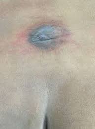
Limited dorsal myeloschisis
Limited dorsal myeloschisis (LDM) is a distinctive form of spinal dysraphism characterized by 2 constant features: a focal “closed” midline defect and a fibroneural stalk that links the skin lesion to the underlying cord.
Embryogenesis
Embryogenesis is hypothesized to be incomplete disjunction between cutaneous and neural ectoderms, thus preventing complete midline skin closure and allowing persistence of a physical link (fibroneural stalk) between the disjunction site and the dorsal neural tube.
However, clinical experience of LDM located below the first-second sacral (S1-S2) vertebral level, which is formed from secondary neurulation (S2-coccyx), suggested that LDM may not be entirely explained as an error of primary neurulation.
Treatment
Its treatment is a relatively straightforward resection of the LDM stalk from the spinal cord.
Reviewing series of 75 cases of LDMs, we found that the majority of LDM stalks have only a glioneuronal core within a fibrous stroma, but a small number have been found to have elements of dermoid cyst or a complete dermal sinus tract either contiguous with the fibroneural stalk or incorporated within its glial matrix, not surprising considering the original continuum of cutaneous and neural ectoderm in LDMs' embryogenesis. The dermoid element can be microscopic and escape casual observation, but could grow to large intradural dermoid cysts if part of the dermoid invested LDM stalk is left inside the dura.
They recommend excising the entire length of the intradural LDM stalk from its dural entry point to its merge point with the spinal cord during the initial treatment to avoid secondary deterioration and additional surgery 1).
Case series
In 14 LDM patients, 2 had tail-like appendages. We retrospectively analyzed the relationship between the appendage and the LDM tract from the clinicopathological findings of these 2 patients.
Preoperative magnetic resonance imaging including three-dimensional heavily T2-weighted images demonstrated an intradural tethering tract, but failed to reveal the precise communication with the appendage. However, surgery revealed the extradural and intradural slender stalk, starting at the base of appendage and running through the myofascial defect. Histological examination demonstrated that there was a tight anatomical relationship between the fibroadipose tissue of the appendage and the fibrocollagenous LDM stalk.
When there is potential for an LDM stalk in patients with an appendage, a meticulous exploration of the stalk leading from an appendage is required. Clinicians should be aware of possible morphological variations of skin lesions associated with LDM 2).
Twenty-eight patients were surgically treated for LDM from 2010 to 2015. Since the level where the LDM stalk penetrates the interspinous ligament is most clearly defined on the preoperative MRI and operative field, this level was assessed to find out whether the lesions can occur in the region of secondary neurulation.
Eleven patients (39%) with typical morphology of the stalk had interspinous defect levels lower than S1-S2. These patients were not different from 17 patients with classic LDMs at a level above or at S1-S2. This result shows that other than the low level of the interspinous level, 11 patients had lesions that could be defined as LDMs.
By elucidating the location of LDM lesions (in particular, the interspinous level), we propose that LDM may be caused by errors of secondary neurulation. The hypothesis seems more plausible due to the supportive fact that the process of separation between the cutaneous and neural ectoderm is present during secondary neurulation. Hence, incomplete disjunction of the two ectoderms during secondary neurulation may result in LDM, similar to the pathomechanism proposed during primary neurulation 3).
Morioka et al., retrospectively analyzed the histopathological findings of the almost entire stalk and their relevance to the clinical manifestations in six Japanese LDM patients with flat skin lesions.
Glial fibrillary acidic protein (GFAP)-immunopositive neuroglial tissues were observed in three of the six patients. Unlike neuroglial tissues, peripheral nerve fibers were observed in every stalk. In four patients, dermal melanocytosis, “Mongolian spot,” was seen surrounding the cigarette-burn lesion. In three of these four patients, numerous melanocytes were distributed linearly along the long axis of the LDM stalk, which might represent migration of melanocytes from trunk neural crest cells during formation of the LDM stalk.
Immunopositivity for GFAP in the LDM stalk was observed in as few as 50% of our patients, despite the relatively extensive histopathological examination. We confirm that the clinical diagnosis of LDM should be made based on comprehensive histopathological examination as well as clinical manifestations. The profuse network of peripheral nerve fibers in every stalk and the high incidence of melanocyte accumulation associated with dermal melanocytosis might assist the histopathological diagnosis of LDM 4).
In 51 LDM patients. all patients were studied with magnetic resonance imaging or computed tomography myelography, operated on, and followed for a mean of 7.4 years.
There were 10 cervical, 13 thoracic, 6 thoracolumbar and 22 lumbar lesions. Two main types of skin lesion were saccular (21 patients), consisting of a skin-base cerebrospinal fluid sac topped with a squamous epithelial dome, and nonsaccular (30 patients), with a flat or sunken squamous epithelial crater or pit. The internal structure of a saccular LDM could be a basal neural nodule, a stalk that inserts on the dome, or a segmental myelocystocele. In nonsaccular LDMs, the fibroneural stalk has variable thickness and complexity. In all LDMs, the fibroneural stalk was tethering the cord. Twenty-nine patients had neurological deficits. There was a positive correlation between neurological grade and age, suggesting progression with chronicity. Treatment consisted of detaching the stalk from the cord. Most patients improved or remained stable.
LDM is a distinctive clinicopathological entity and a tethering lesion with characteristic external and internal features. We propose a new classification incorporating both saccular and flat lesions 5).
