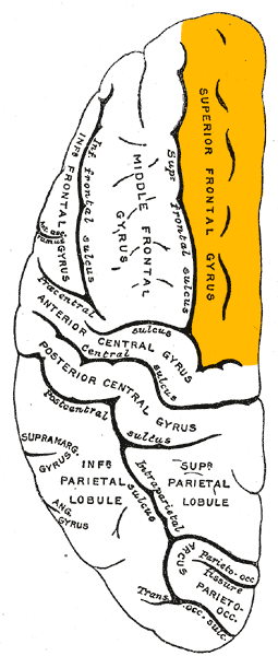
Left middle frontal gyrus
Results suggest that, compared to patients with a intracranial meningioma at other locations, patients with a meningioma at the left middle frontal gyrus are at potential risk for worse performance on cognitive flexibility and complex attention whereas patients with a meningioma at the left superior frontal gyrus are at potential risk for worse performance on complex attention 1).
To assess the specific roles of left middle frontal gyrus (LMFG) in word production, electrocorticography signals were recorded from an epilepsy patient when he participated in language tasks. Wen et al. found three sites of LMFG showed high-gamma perturbations with distinct patterns across tasks; and neural activities elicited in the same tasks shared similar patterns, while those elicited by stimuli leading to the same articulations did not. These findings confirmed that the LMFG takes active parts in word production, and suggested that it may serve as a temporal perceptual information storage space, supporting the hierarchical state feedback control model of word production 2).
In 2009 Andersson et al. suggested that the left middle frontal gyrus is a part of an executive attention network, and that the dichotic listening forced attention paradigm may be a feasible tool for assessing subtle attentional dysfunctions in older adults 3).
A 65-year-old right-handed man noted a sudden onset of numbness and weakness of the right hand. On the initial visit to the hospital, he showed severe acalculia, and transient agraphia (so called incomplete Gerstmann syndrome) and transcortical sensory aphasia. Brain MRI revealed a fresh infarct in the left middle frontal gyrus. The paragraphia and aphasia improved within 14 days after onset, but the acalculia persisted even at seven months after onset In an 123I-IMP SPECT study, the cerebral blood flow (CBF) was found to be decreased in the infarction lesion and its adjacent wide area, the ipsilateral angular and supramarginal gyri, and contralateral cerebellar hemisphere. Ando et al., speculate that inactivation in the infarction lesion caused the CBF decrease in the non-infarcted areas due to diaschisis. This case indicates that Gerstmann syndrome can be caused by not only dysfunction of the angular gyrus but also of the left middle frontal gyrus in the dominant hemisphere 4).
