Indocyanine green videoangiography indications for vascular neurosurgery
Indocyanine green (ICG) fluorescence videoangiography is now widely used for the intraoperative assessment of vessel patency, providing high quality, valuable, real-time imaging of cerebrovascular anatomy.
Preservation of adequate blood flow and exclusion of flow from lesions are key concepts of vascular neurosurgery.
The overlay of fluorescence videoangiography within the field of view of the white light operative microscope allows real-time assessment of the blood flow within vessels during simultaneous surgical manipulation. This technique could improve intraoperative decision making during complex neurovascular procedures 1).
Simal-Julián et al. conducted a review to identify and assess the impact of all of the methodological variations of conventional ICGVA applied in the field of neurovascular pathology that have been published to date in the English literature. A total of 18 studies were included in this review, identifying four primary methodological variants compared to conventional ICGVA: techniques based on the transient occlusion, intra-arterial ICG administration via catheters, use of endoscope system with a filter to collect florescence of ICG, and quantitative fluorescence analysis. These variants offer some possibilities for resolving the limitations of the conventional technique (first, the vascular structure to be analyzed must be exposed and second, vascular filling with ICG follows an additive pattern) and allow qualitatively superior information to be obtained during surgery. Advantages and disadvantages of each procedure are discussed. More case studies with a greater number of patients are needed to compare the different procedures with their gold standard, in order to establish these results consistently 2).
It can assist in intraoperative surgical management and/or stroke prevention particularly during aneurysm clipping, EC-IC bypass and AVM/DAVF surgery 3), and to document the intraoperative vascular flow 4).
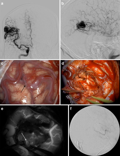
Indocyanine green (ICG) angiography is commonly used to map the vascular configuration of cerebral arteriovenous malformations (AVMs) during resection.
ICG-VA is a safe and effective technique for locating the ICA in skull-base expanded endonasal surgery. Furthermore, this technique can provide real-time guidance for the surgeon and increase safety for the patient 5).
Vascular malformations
Sato et al. developed a new, high-resolution intraoperative imaging system (dual-image VA [DIVA]) to simultaneously visualize both light and near-infrared (NIR) fluorescence images from ICG-VA, allowing observation of other structures.
The operative field was illuminated via an operating microscope by halogen and xenon lamps with a filter to eliminate wavelengths over 780 nm. In the camera unit, visible light was filtered to 400-700 nm and NIR fluorescence emission light was filtered to 800-900 nm using a special sensor unit with an optical filter. Light and NIR fluorescence images were simultaneously visualized on a single monitor.
The system clearly visualized the operative field together with fluorescence-enhanced blood flow. In aneurysm surgeries, we could confirm incomplete clipping with the neck remnant or with remnant flow into the aneurysm. In cases of arteriovenous malformation or arteriovenous fistula, feeding arteries and draining veins were easily distinguished.
This system allows observation of the operative field and enhanced blood flow by ICG together in real-time and may facilitate various types of neurovascular surgery 6).
