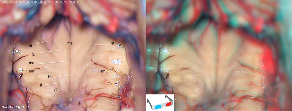
Fourth ventricle floor
The floor (i.e. the anterior edge) of the fourth ventricle constitutes the rhomboid fossa, and comprises a number of general features.
A sulcus - the median sulcus - extends the length of the ventricle (from the cerebral aqueduct of the midbrain to the central canal of the spinal cord), dividing the floor into right and left halves. Each half is further divided by a further sulcus - the sulcus limitans - along a line parallel to the median sulcus; within the floor, motor neurons are located medially of the sulcus limitans, while sensory neurons are located laterally. The elevation between the median sulcus and sulcus limitans (i.e. the region for motor neurons), is known as the medial eminence, while the lateral region (i.e. that for the sensory neurons) is known as the vestibular area. The sulcus limitans bifurcates at either end - the superior fovea cerebrally, and the inferior fovea caudally.
The posterior surface of the pons and the upper open part of the medulla oblongata form the floor of the fourth ventricle. It is covered by ependyma and because of its rhomboid shape, is called the rhomboid fossa.
Floor of the fourth ventricle: fc: facial colliculus; ht: hypoglossal triangle; if: inferior fovea; me: medial eminence; ms: median sulcus; o: obex; sf: superior fovea; sl: sulcus limitans; sm: medullary striae of fourth ventricle; vt: vagal triangle.
The lower end of the fossa is called the obex; the upper end continues into the aqueduct of the midbrain.
The lateral elevation, also called the vestibular area, is produced largely by the underlying vestibular nuclear complex. The sulcus limitans demarcates afferent and efferent cell columns. A number of white strands, called striae medullares, run across the caudal part of the rhomboid fossa (i.e., the medullary part, hence the name striae medullares). They are most probably fibres on their way to the cerebellum.
Studies have used the Twining line (from the tuberculum sellae to the torcula), or the floor of the fourth ventricle as reference lines and defined different cutoff values of “steepness”. The other important factors to be considered in approaching the pineal region are anatomic features of the quadrigeminal cistern and superior cerebellar cistern, as both host important neurovascular structures and serve as anatomic corridors 1) 2) 3) 4) 5).
The current trend to use the Twining line to define this angle has significant pitfalls.

