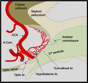
Subcallosal artery
The subcallosal artery is a unique vessel because it supplies the septal/subcallosal region bilaterally; preservation of this vessel during surgery is crucial for successful outcomes 1).
During surgery to treat an anterior communicating artery aneurysm, injury to the subcallosal artery, a perforator of the anterior communicating artery, may lead to infarction that produces basal forebrain amnesia after surgery.
Despite the variable anatomy of the anterior communicating artery (AcoA) complex, three main perforating branches can be typically identified the largest of which being the subcallosal artery (ScA).
In 33 specimens from transcranial and endonasal perspectives, the ScA was present in 79% of the specimens as a single dominant artery arising from the posterior/posterosuperior surface of the AcomA, along with hypothalamic arteries (55%), or as a single artery (24%). It coursed posteriorly towards the lamina terminalis region, curving superiorly to the subcallosal area. The ScA gave off many branches to provide the main blood supply to the subcallosal region. Importantly, it supplies the septal/subcallosal region bilaterally. The ScA can be found posterior, superior, or inferior to the AcomA when using a transylvian, interhemispheric, or endonasal approach, respectively. In specimens with no ScA (21%), the median callosal artery (MdCA) was the dominant artery arising from the AcomA. It followed an identical course to the ScA, providing supply to the same structures bilaterally, but its distal extension reached the body/splenium of the corpus callosum. The MdCA is a ScA variant 2).
In 30 cadaver brains, the ACoA and its branches were examined under magnification using a surgical microscope.
The ACoA was evident in all specimens and had variations consisting of plexiform (33%), dimple (33%), fenestration (21%), duplication (18%), string (18%), fusion (12%), median artery of the corpus callosum (6%), and azygous anterior cerebral artery (3%). The perforating branches were also observed in all cadaver brains. They were classified into subcallosal, hypothalamic, and chiasmatic branches according to their vascular territories. The subcallosal branch, usually single and the largest, supplied the bilateral subcallosal areas, branching off to the hypothalamic area. The hypothalamic branches, multiple and of small caliber, terminated in the hypothalamic area.
The incidence of anomalous ACoA was higher than has been previously reported, and any segment of the anomalous ACoA may have perforating branches regardless of diameter.
Despite the variable anatomy of the anterior communicating artery (AcoA) complex, three main perforating branches can be typically identified the largest of which being the subcallosal artery (ScA).
This artery is very important because it feeds bilateral subcallosal areas branching to the hypothalamic area 3).
Stroke in the vascular territory of the ScA leads to a characteristic imaging and clinical pattern. Ischemia involves the anterior columns of the fornix and the genu of the corpus callosum, and patients present with a Korsakoff's syndrome including disturbances of short-term memory and cognitive changes. We conclude that despite its small size, the ScA is an important artery to watch out for during surgical or endovascular treatment of AcoA aneurysms 4).
Mugikura et al., evaluated 3D-T2-weighted MR images obtained a median of 4 months after treatment of anterior communicating artery aneurysm for the presence of infarcted foci in 10 consecutive patients with postoperative amnesia. Because the subcallosal artery and its neighboring perforator, the recurrent artery of Heubner, were considered the most easily affected vessels during that surgery, they focused mainly on 8 regions of the subcallosal artery territory per hemisphere and 5 regions of the recurrent artery of Heubner territory per hemisphere.
All 10 patients had infarcts in the territory of the subcallosal artery (median, 9 regions per patient), and most were bilateral (9 of 10 patients). Five patients had additional infarcted foci in the territory of the recurrent artery of Heubner (median, 1 region per patient), all unilateral. Among the regions perfused by the subcallosal artery, the column of the fornix was involved in all patients; the anterior commissure, in 9; and the paraterminal gyrus, in 8 patients.
3D MR imaging revealed subcallosal artery infarction, the distribution of which was mostly bilateral, presumably owing to the unpairedness of that artery, in patients with postoperative amnesia after anterior communicating artery aneurysm repair 5).
