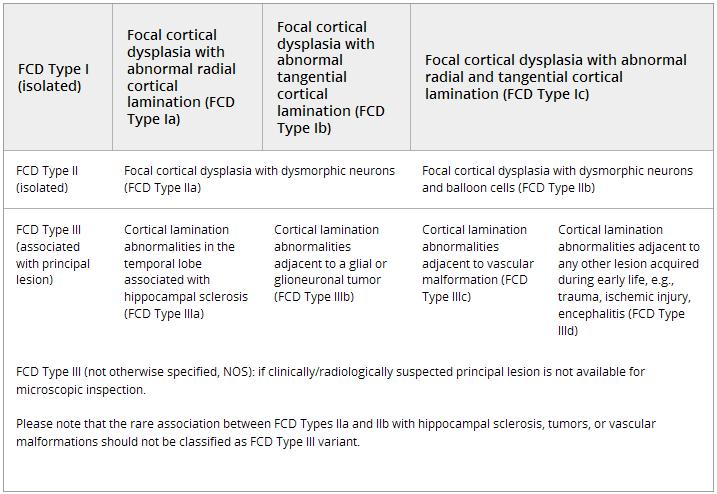
Focal cortical dysplasia classification
The Diagnostic Methods commission of the International League against Epilepsy (ILAE) released a first international consensus classification of Focal Cortical Dysplasia (FCD) in 2011. Since that time, this FCD classification has been widely used in clinical diagnosis and research (more than 740 papers cited in Pubmed between 1/1/2012 and 7/1/2017).
see Focal cortical dysplasia type I
see Focal cortical dysplasia type II.
see Focal cortical dysplasia type II B.
see Focal cortical dysplasia type III
see Focal cortical dysplasia type IIIB.
International League Against Epilepsy (ILAE) classifications of FCD.
Results suggest that the current FCD classification should recognize a panel of immunohistochemical stainings for a better histopathological workup and definition of FCD subtypes. Blümcke et al. also proposed adding the level of genetic findings to obtain a comprehensive, reliable, and integrative genotype-phenotype diagnosis in the near future 1).
Focal Cortical Dysplasia types I, II, and III, are associated with somatic gene variants across a broad range of genes, many associated with epilepsy in clinical syndromes caused by germline variants, as well as including some not previously associated with radiographically evident cortical brain malformations 2).
Previous studies have suggested that alteration of cortical interneurons and abnormal cytoarchitecture have been linked to initiation and development for seizure. However, whether each individual subpopulation of cortical interneurons is linked to distinct FCD subtypes remains largely unknown.
Liang et al. retrospectively analyzed both control samples and epileptic specimens pathologically diagnosed with FCD types Focal cortical dysplasia type IA, Focal cortical dysplasia type IIA, or Focal cortical dysplasia type IIB.
They quantified three major interneuron (IN) subpopulations, including parvalbumin (PV)-, somatostatin (Sst)-, and vasoactive intestinal peptide (Vip)-positive INs across all the subgroups. Additionally, they calculated the ratio of the subpopulations of INs to the major INs (mINs) by defining the total number of the PV-, Sst-, and Vip-INs as mINs. Compared with the control, the density of the PV-INs in FCD type IIb was significantly lower, and the ratio of PV/mINs was lower in the superficial part of the cortex of the FCD type Ia and IIb groups. Interestingly, we found a significant increase in the ratio of Vip/mINs only in FCD type IIb. Overall, these results suggest that in addition to a reduction in PV-INs, the increase in Vip/mINs may be related to the initiation of epilepsy in FCD type IIb. Furthermore, the increase in Vip/mINs in FCD type IIb may, from the IN development perspective, indicate that FCD type IIb forms during earlier stages of pregnancy than FCD type Ia 3).
