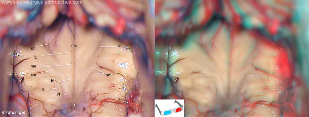
Facial colliculus
The facial colliculus is an elevated area located on the dorsal pons in the floor of the fourth ventricle. It is formed by fibers from the motor nucleus of the facial nerve as they loop over the abducens nucleus. Thus a lesion to the facial colliculus would result in ipsilateral facial nerve paralysis and ipsilateral unopposed eye medial deviation.
Fourth ventricle floor: fc: facial colliculus; ht: hypoglossal triangle; if: inferior fovea; me: medial eminence; ms: median sulcus; o: obex; sf: superior fovea; sl: sulcus limitans; sm: medullary striae of fourth ventricle; vt: vagal triangle.
The facial colliculus (FC), an important landmark for planning a surgical brainstem cavernous malformation approach (BCM), it may be difficult to identify on magnetic resonance imaging (MRI). Three-dimensional (3D) images may improve the FC-identification certainty; hence, a study attempted to validate the FC-identification certainty between two-dimensional (2D) and 3D images of patients with a normal brainstem and those with BCM. In this retrospective study, Uchida et al. included 10 patients with a normal brainstem and 10 patients who underwent surgery for BCM. The region of the FC in 2D and 3D images were independently identified by three neurosurgeons, three times in each case, using the method for continuously distributed test results (0-100). The intra- and inter-rater reliability of the identification certainty was confirmed using the intraclass correlation coefficient (ICC). The FC-identification certainty for 2D and 3D images was compared using the Wilcoxon signed-rank test. The ICC (1,3) and ICC (3,3) in both groups ranged from 0.88 to 0.99; therefore, the intra- and inter-rater reliability were good. In both groups, the FC-identification certainty was significantly higher for 3D images than for 2D images (normal brainstem group; 82.4 vs. 61.5, P = .0020, BCM group; 40.2 vs. 24.6, P = .0059 for the unaffected side, 29.3 vs. 17.3, P = .0020 for the affected side). In the normal brainstem and BCM groups, 3D images had better FC-identification certainty. 3D images are effective for the identification of the FC 1).
