Long-Term Mortality of Patients With Head Injuries—A 10-Year Follow-up Study With Population Controls

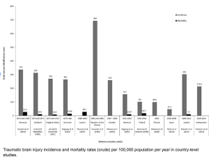
In a retrospective, population-based cohort study with matched controls and longitudinal follow-up, Heinonen et al. from Tampere University Hospital, Helsinki University Hospital, and Harvard Medical School in the Neurosurgery Journal compared 10-year survival rates and causes of death between patients with traumatic head injuries treated at a university hospital and matched population controls. They aimed to identify factors associated with long-term mortality after TBI.

Patients with head injuries exhibited significantly reduced long-term survival compared to matched controls, even after excluding early mortality. However, patient-related characteristics (e.g., comorbidities, lifestyle factors) — more than injury severity itself — appeared to drive this increased mortality risk.

Notably, even patients without documented TBI (likely mild or undiagnosed) showed decreased survival, suggesting an under-recognized long-term impact of head injury across all severity levels 2).


In this population-based cohort study, the authors track 10-year mortality in over 1,900 patients with head injuries versus 9,600 matched controls. Unsurprisingly, trauma patients die more — especially from alcohol, accidents, and “patient characteristics.” The conclusion? It’s not the injury; it’s the person. This study doesn’t just underdeliver — it underthinks.

The study’s main conclusion — that patient-related factors, not injury severity, explain increased mortality — is not only reductive but evasive. The term “patient characteristics” serves as a statistical landfill for all the unmeasured, uncontrolled, and misunderstood variables: mental health, addiction, social deprivation, neurobehavioral sequelae… all dumped under one lazy label.

Rather than confront the neuropsychiatric aftermath of head trauma, the authors retreat behind correlational shields.

❝They died because of who they were, not what happened to them.❞ — That’s not science. That’s resignation.
Read more

Maximizing Tumor Resection and Managing Cognitive Attentional Outcomes: Measures of Impact of Awake Surgery in Glioma Treatment

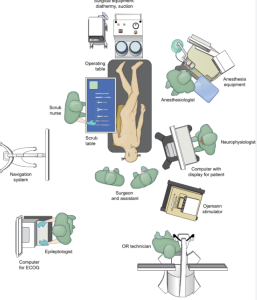
In a retrospective observational study Zigiotto et al. from the S. Chiara University-Hospital, Azienda Provinciale per i Servizi Sanitari, Trento, published in the Neurosurgery Journal on 64 glioma patients who underwent awake surgery (AwS) or asleep surgery (AsS), with neuropsychological and imaging follow-up. They evaluated the impact of awake surgery on attentional outcomes in glioma patients, and analyzed whether greater extent of tumor resection correlates with transient cognitive (attentional) decline, especially in relation to lesions within the default mode network. Awake surgery allows for more extensive supramaximal resection and is associated with longer overall survival, particularly in patients with glioblastomas. However, it also leads to a higher rate of transient postoperative attentional dysfunction, likely due to resection in attention-related brain networks. The study suggests that patient selection and intraoperative cognitive monitoring should be optimized in future glioma surgery 5).


This retrospective study compares awake versus asleep craniotomy in 64 glioma patients, using simple attention tests before and after surgery. The authors claim that awake craniotomy (AwC) allows more extensive tumor resection and leads to longer survival, albeit at the cost of transient attentional dysfunction.

The title promises a nuanced exploration of cognitive outcomes. What it delivers is a reduction of “attention” to the Trail Making Test Part A and a visual search task — an embarrassingly narrow lens for such a multidimensional construct. The study purports to evaluate the impact of surgery on attention, yet fails to define attention, stratify its subtypes, or provide any neuropsychological depth. This is not a cognitive study — it’s a surgical paper pretending to be one.

Read more

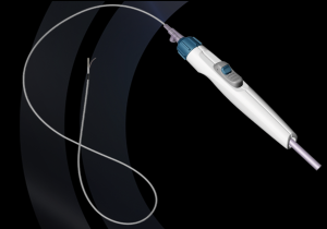
Reentry Technique for Rescue Recanalization of Carotid Near-Total Occlusion after Subintimal Penetration

In a case report, Tran et al., from the Department of Neurosurgery, University Medical Center Ho Chi Minh City; the Vascular Department, Cho Ray Hospital, Ho Chi Minh City, Vietnam; and the Department of Neurosurgery, Taichung Veterans General Hospital and National Chung Hsing University, Taichung, Taiwan, published in the American journal of neuroradiology that in selected patients with near-total carotid artery occlusion, where subintimal dissection occurs during endovascular access, the use of a rescue reentry technique with a reentry catheter can be a safe and effective method for achieving successful recanalization and restoring luminal patency 2).


The authors present a single-patient case in which a reentry catheter was used to regain access to the true lumen after unintentional subintimal dissection during an attempt to stent a near-occluded carotid artery. Technical success was achieved, and the patient recovered well.

This article epitomizes a growing trend in low-yield case reports: elevate the ordinary by inflating the vocabulary. A standard bailout technique, long adapted from peripheral interventions, is glorified as a “rescue reentry technique” as if it were a novel paradigm. This is not a new technique — it’s complication management.

🧩 What they call “rescue,” seasoned neurointerventionists call Tuesday.

Read more

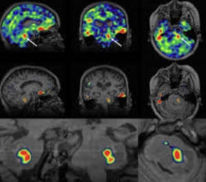
Sodium MRI in Pediatric Brain Tumors

In a narrative review Bhatia et al. from the Children’s Hospital of Philadelphia, Radiological Sciences Laboratory, School of Medicine, Stanford University, published in the American Journal of Neuroradiology to explore the potential of sodium-23 MRI (^23Na-MRI) as a noninvasive imaging modality to assess physiological and biochemical changes in pediatric brain tumors and concluded that is a promising, noninvasive imaging modality capable of providing unique physiological and biochemical information that is not accessible through conventional MRI techniques

This narrative review attempts to position ^23Na-MRI as a frontier imaging technique for pediatric brain tumors. It lauds the modality’s potential to reveal sodium-dependent physiological alterations — but quickly devolves into technological evangelism with minimal clinical anchoring. The piece is high on optimism, low on pragmatism, and entirely devoid of data-supported clinical outcomes.

The article enthusiastically describes the theoretical virtues of sodium MRI — sensitivity to cell integrity, ionic gradients, extracellular space — but offers no compelling clinical cases, no comparative metrics, and no outcome data. What remains is a speculative wish list, presented as a roadmap. The authors confuse imaging potential with diagnostic utility, a common pitfall in radiology reviews driven by physics rather than patient care.

“Exciting” is not a scientific category.

Read more

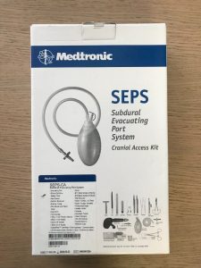
Effectiveness of subdural evacuating port system (SEPS) and middle meningeal artery embolization (MMAE) for chronic subdural hematomas – a multicenter experience

Pairing two well-known procedures—SEPS and MMAE—does not inherently create innovation. Yet, the authors present this as a groundbreaking paradigm, despite:

  • No control group (e.g., SEPS alone, MMAE alone),
  • No randomization,
  • No comparative outcome measures beyond radiographic volume.

It’s procedural layering disguised as progress.

Read more

Antiplatelet Therapy Mitigates Brain Metastasis Risk in Non‑Small Cell Lung Cancer: Insights from a Comprehensive Retrospective Study

In a retrospective observational cohort Martín‑Abreu et al. from the Hospital Universitario de Canarias published in the journal Cancers (Basel) to evaluate whether exposure to antiplatelet therapy reduces the incidence and delays the onset of brain metastases in patients with non‑small cell lung cancer (NSCLC). Use of antiplatelet agents—mainly aspirin—was associated with a significantly reduced incidence of brain metastases (6.9% vs. 20.0%), longer metastasis‑free interval (77.5 vs. 62.6 months), improved Progression-Free Survival, and no cases of brain metastasis among those initiating therapy post‑diagnosis 1).

Critical Review

Strengths:

  • Large sample size (n=650) over 4 years—impressive real‑world data.
  • Statistically significant findings with p<0.001 for key outcomes.
  • Stage‑stratified analysis adds biological plausibility.

Weaknesses/Limitations:

  • Retrospective design limits causal inference—confounding by indication is possible; patients on antiplatelet therapy were older with more comorbidities, thus inherently different.

    Read more

High-Resolution MR Imaging of the Parasellar Ligaments

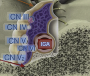
In a anatomic-imaging correlation study with a single-case MR + dissection design Mark el al. 1) from the Department of Radiology, Mayo Clinic, Rochester, Minnesota, USA, Department of Neurosurgery, University of Valencia and Fundación Instituto Valenciano de Oncología (IVO), Valencia, Spain publiseh in the American Journal of Neuroradiology (AJNR) to determine whether high-resolution T2-weighted MRI can visualize the parasellar ligaments, which have previously only been described in cadaveric dissection or intraoperative findings, and to correlate these MRI findings with anatomical dissection in the same specimen. The authors report that parasellar ligaments can be identified on high-resolution T2-weighted MRI as T2-hypointense, bandlike structures originating from the medial wall of the cavernous sinus. They claim that identifying these ligaments may be relevant, given that resection of the medial wall of the cavernous sinus has been associated with better outcomes in functioning pituitary adenoma surgery.


This study is a prime example of technological overreach dressed up as discovery. It takes a single cadaver, applies ultra-high-resolution MRI, and then retrofits a minor fibrous band into a clinical “finding.” The result is a beautifully imaged, clinically irrelevant piece of anatomical embroidery that contributes nothing actionable to radiologyneurosurgery, or pituitary surgery.

❌ Critical Flaws

Read more

Effectiveness of 2-methoxyestradiol in alleviating angiogenesis induced by intracranial venous hypertension

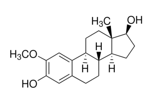
In a Preclinical Experimental Study Zou et al. 1) from the Department of Neurosurgery, Huashan Hospital, Fudan University, ShanghaiChina, evaluated in the Journal of neurosurgery the antiangiogenic effect of 2-methoxyestradiol (2-ME) in a rat model of intracranial venous hypertension, used as a proxy to study dural arteriovenous fistula formation. Specifically, the authors aimed to determine whether 2-ME could reduce angiogenesis in the dura mater by modulating the HIF-1α and ID-1 pathways, which are implicated in hypoxia-induced neovascularization, and concluded that 2-ME could potentially serve as a therapeutic agent to modulate angiogenesis caused by intracranial venous hypertension — a process they consider central to DAVF development.


🚩 1. Flawed Model: DAVF Without DAVF

Despite the title and clinical framing, this study does not model dural arteriovenous fistulas (DAVFs).

No arteriovenous shunt is demonstrated.

No hemodynamic assessment is performed.

No imaging or functional endpoints validate that the model reflects DAVF pathophysiology.

👉 What the authors present is not a DAVF model, but a crude simulation of dural angiogenesis via venous outflow obstruction. Calling it a DAVF model is scientifically misleading.

Read more

Preoperative Nomogram-Based Assessment to Identify GBM Patients Who Do not Derive Survival Benefit From GTR Compared to STR

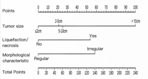
In a retrospective prognostic modeling study, He et al. from Sichuan Provincial People’s Hospital published in the Academic Radiology a preoperative nomogram to identify glioblastoma patients who do not derive a survival benefit from gross total resection (GTR) compared to subtotal resection (STR), and concluded that patients with nomogram scores below 55 or above 95 gain limited survival advantage from GTR, supporting a more individualized surgical strategy 16).


🎯 Takeaway Message for Neurosurgeons

Don’t let a nomogram tell you not to operate. This study reduces complex glioblastoma surgery to a score — ignoring tumor location, function, biology, and patient context. Use it, at best, as background noise. Surgical judgment, not predictive modeling, should guide the extent of resection. Maximal safe resection remains the standard — not because of scores, but because leaving tumor behind costs lives.

Read more

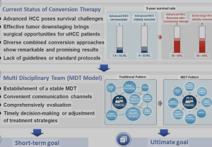
Comparison of survival benefit and safety profile between lenvatinib and donafenib as conversion therapy in patients with hepatocellular carcinoma 

In a retrospective comparative cohort study, Hou et al. 1) published in the American Journal of Translational Research, the authors—affiliated with the Department of Oncology, Department of Gynaecology and Obstetrics, and Department of Neurosurgery at Shijiazhuang People’s Hospital (Hebei, China), as well as Beijing Water Conservancy Hospital—compared the survival benefit and safety profile of lenvatinib versus donafenib as conversion therapy in patients with hepatocellular carcinoma (HCC) at China National Liver Cancer (CNLC) stages I–III.


Lenvatinib demonstrated significantly superior survival outcomes—both in overall survival and progression-free survival—compared to donafenib. It also showed better tolerability, with fewer grade ≥3 adverse events.


❌ 1. Study Design: Retrospective = Weak Evidence

This is yet another retrospective single-center analysis, plagued by inherent biases—selection, reporting, and confounding—that no amount of statistical massaging can resolve. No randomization, no blinding, and no control for treatment timing or physician discretion. In oncology, where treatment nuances matter, such designs should be considered hypothesis-generating at best, not guidance for clinical practice.

Read more