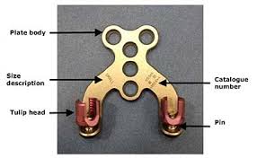
Occipital plate
In 1999, Pait et al. described the “inside-outside” technique, in which a screw with a long plating system is connected from the occipital bone down to the posterior cervical elements 1). The construct was intended to decrease screw pullout because the suboccipital portion of the bone has an inverted screw placed into a burr hole. Vale et al. attempted to decrease screw pullout with a similar system that utilizes a “T-plate” device 2). In place of rods, an elongated plate attaches to the occipital keel superiorly and to the lateral mass in the spine. One of the most common titanium hardware systems included a Y-shaped plating system that was adopted from orthopaedic long-bone fractures.
After lateral intraoperative images confirm alignment of the CCJ, the plate is centered at the occipital keel. In the authors’ technique, once the proposed placement of the occipital plate is finalized, pilot holes for the occipital screws through the cortical bone are made using a high-speed matchstick burr. Then, using a power drill, the rest of the screw path is cannulated with the use of a drill guide. Preoperative computed tomography scans can be helpful for planning and sizing appropriate screw length. Depending on the patient anatomy, we advocate at least a 10-mm length screw, and often 12- or 14-mm screws can be used. A ball-tipped feeler can be used to palpate the bottom of the hole to see if there is cortical bone remaining versus dura. In the event of an unintended durotomy with visible egress of cerebral spinal fluid (CSF), no additional repair is required beyond placement of the screw. Bone wax can be used to temporize the leak if there is significant flow which may obscure the surgical field. Maximizing screw length in the occipital keel maximizes bony purchase and ensures construct rigidity. In the pediatric population, there is often times a persistent dural venous sinus in the midline. It is of the utmost importance to avoid lacerating this structure as significant bleeding can result. Careful preoperative planning in such cases is paramount to avoiding such surgical morbidity. In addition, most modern occipital plates have lateral screw holes available to stabilize against rotation forces on the occipital plate. The thickness of the occipital bone here is thinner compared to the keel, and we recommend 6- to 8-mm length screws.
Once the size of the occipital plate screws has been confirmed, the hole is tapped and the screw is inserted. While initial constructs consisted of an occipital bone plate connected to the cervical rods, modern plates now have screws on the lateral plate to accommodate pre-bent or jointed rods. We do not recommend the bending of a straight rod to accommodate the occipitocervical junction as this contributes to metal fatigue, and the gap between the occiput and the most proximal cervical fixation is an area highly susceptible to rod fracture. The distal aspect of the rod must be affixed with posterior cervical spine screws, which in some cases can lead to difficulty with rod placement. Our protocol is to place the occipital plate first, which allows for alignment of distal screws in line to facilitate rod capture.
Proximal stabilization is usually ensured by bi-cortical occipital screws implanted through one median or two lateral occipital plate(s). Bone thickness variability as well as the proximity of vasculo-nervous elements can induce substantial morbidity. The choice of site and implant type remains difficult for surgeons and is often empirically based. Given this challenge, implants with smaller pitch to increase bone interfacing are being developed, as is a surgical technique consisting in inverted occipital hook clamps, a potential alternative to plate/screws association.
Germaneau et al. presented a biomechanical comparison of the different occipitocervical fusion devices.
They developed a 3D mark tracking technique to measure experimental mechanical data on implants and occipital bone. Biomechanical tests were performed to study the mechanical stiffness of the occipito-cervical instrumentation on human skulls. Four occipital implant systems were analysed: lateral plates+large pitch screws, lateral plates+hooks, lateral plates+small pitch screws and median plate+small pitch screws. Mechanical responses were analysed using 3D displacement field measurements from optical methods and compared with an analytical model.
Paradoxical mechanical responses were observed among the four types of fixations. Lateral plates+small pitch screws appear to show the best accordance of displacement field between bone/implant/system interface providing higher stiffness and an average maximum moment around 50 N.m before fracture.
Interpretation: Stability of occipito-cervical fixation depends not only on the site of screws implantation and occipital bone thickness but is also directly influenced by the type of occipital implant 3).
