Middle temporal gyrus
The middle temporal gyrus is located between the superior temporal gyrus and inferior temporal gyrus.
The middle temporal gyrus is bounded by:
the superior temporal sulcus above;
the inferior temporal sulcus below; an imaginary line drawn from the preoccipital notch to the lateral sulcus posteriorly.
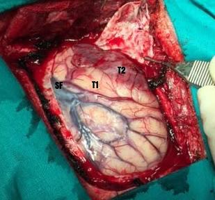
The bone flap has been removed and the dura mater has been opened as a flap pediculated towards the greater sphenoid wing previously roungered to improve parasellar visualization. Sylvian fissure, Inferior frontal gyrus, Superior temporal gyrus and Middle temporal gyrus are exposed. Three pars of parasylvian inferior frontal gyrus must be distinguished: pars orbitalis (pOr) in relation to the orbital roof; pars triangularis (pT) the widest area of sylvian fissure (good place for start opening of sylvian fissure); pars opercularis (pOp) where Broca’s Area is located.
Approaches
Functions
The middle temporal gyrus (MTG) participates in a variety of functions, suggesting the existence of distinct functional subregions. In order to further delineate the functions of this brain area, Xu et al., parcellated the MTG based on its distinct anatomical connectivity profiles and identified four distinct subregions, including the anterior (aMTG), middle (mMTG), posterior (pMTG), and sulcus (sMTG). Both the anatomical connectivity patterns and the resting-state functional connectivity patterns revealed distinct connectivity profiles for each subregion. The aMTG was primarily involved in the default mode network, sound recognition, and semantic retrieval. The mMTG was predominantly involved in the semantic memory and semantic control networks. The pMTG seems to be a part of the traditional sensory language area. The sMTG appears to be associated with decoding gaze direction and intelligible speech. Interestingly, the functional connectivity with Brodmann's Area (BA) 40, BA 44, and BA 45 gradually increased from the anterior to the posterior MTG, a finding which indicated functional topographical organization as well as implying that language processing is functionally segregated in the MTG. These proposed subdivisions of the MTG and its functions contribute to understanding the complex functions of the MTG at the subregional level 1).
EEG was recorded in twenty-two adults while they were asked to (i) envision future monetary episodes; (ii) wait for rewards and (iii) rest. Activation sources were localized to core DMN regions. EEG power and phase coherence were compared across conditions. Prospection, compared to resting and waiting, was associated with reduced power in the medial prefrontal gyrus and increased power in the bilateral medial temporal gyrus across frequency bands as well as greater phase synchrony between these regions in the delta band. The current quantitative EEG analysis confirms prior fMRI research suggesting that medial prefrontal and medial temporal gyrus interactions are central to the capacity for episodic prospection 2).
Bilateral middle temporal gyrus (MTG) regions, anterior to extrastriate body area and the human middle temporal complex, were involved in the visual evaluation of action rationality. These MTG regions are embedded in the superior temporal sulcus regions processing the kinematics of observed actions. Our results suggest that rationality is assessed initially by purely visual computations, combining the kinematics of the action with the physical constraints of the environmental context. The MTG region seems to be sensitive to the contingent relationship between a goal-directed biological action and its relevant environmental constraints, showing increased activity when the expected pattern of rational goal attainment is violated 3).
Some studies indicate that lesions of the posterior region of the middle temporal gyrus, in the left cerebral hemisphere, may result in alexia and agraphia for kanji characters (characters of Chinese origin used in Japanese writing).
Left middle temporal gyrus
The gray matter volume decrease of the left MTG may be utilized as a candidate biomarker for schizophrenia 4).
Right middle temporal gyrus
Results revealed the gray matter reduction of right MTG and bilateral caudate, and disrupted functional connection to widely distributed circuitry in default-mode network (DMN) and frontal regions, respectively. These results suggest that the abnormal DMN and reward circuit activity might be biomarkers of depression trait 5).
The D amino acid oxidase activator gene (G72) has been found associated with several psychiatric disorders such as schizophrenia, major depression, and bipolar disorder. Impaired performance in verbal fluency tasks is an often replicated finding in the mentioned disorders. In functional neuroimaging studies, this dysfunction has been linked to signal changes in prefrontal and lateral temporal areas and could possibly constitute an endophenotype. Therefore, it is of interest whether genes associated with the disorders, such as G72, modulate verbal fluency performance and its neural correlates. Ninety-six healthy individuals performed a semantic verbal fluency task while brain activation was measured with functional MRI. All subjects were genotyped for two single nucleotide polymorphisms (SNP) in the G72 gene, M23 (rs3918342) and M24 (rs1421292), that have previously shown association with the above-mentioned disorders. The effect of genotype on brain activation was assessed with fMRI during a semantic verbal fluency task. Although there were no differences in performance, brain activation in the right middle temporal gyrus (BA 39) and the right precuneus (BA 7) was positively correlated with the number of M24 risk alleles in the G72 gene. G72 genotype does modulate brain activation during language production on a semantic level in key language areas. These findings are in line with structural and functional imaging studies in schizophrenia, which showed alterations in the right middle temporal gyrus 6).
Posterior middle temporal gyrus
The posterior middle temporal gyrus (MTG) and inferior frontal gyrus (IFG) are two critical nodes of the brain's language network. Previous neuroimaging evidence has supported a dissociation in language comprehension in which parts of the MTG are involved in the retrieval of lexical syntactic information and the IFG in unification operations that maintain, select, and integrate multiple sources of information over time.
Data support the view that both left inferior frontal gyrus (LIFG) and Posterior middle temporal gyrus (pMTG) contribute to picture name retrieval, with both sites playing a critical role in mediating the semantic facilitation of naming when retrieval demands are high 7).
Data indicated that Posterior middle temporal gyrus (pMTG) contributes to the controlled retrieval of conceptual knowledge, while angular gyrus AG is critical for the efficient automatic retrieval of specific semantic information 8).
The situation of dissociative amnesia with disproportionate retrograde amnesia is clinically heterogeneous between individuals. Findings may suggest that impairment of high-level integration of visual and/or emotional information processing involving dysfunction of the right posterior middle temporal gyrus could reduce triggering of multi-modal visual memory traces, thus impeding reactivation of aversive memories 9).
Making sense of the world around us depends upon selectively retrieving information relevant to our current goal or context. However, it is unclear whether selective semantic retrieval relies exclusively on general control mechanisms recruited in demanding non-semantic tasks, or instead on systems specialised for the control of meaning. One hypothesis is that the left posterior middle temporal gyrus (pMTG) is important in the controlled retrieval of semantic (not non-semantic) information; however this view remains controversial since a parallel literature links this site to event and relational semantics. In a functional neuroimaging study, we demonstrated that an area of pMTG implicated in semantic control by a recent meta-analysis was activated in a conjunction of (i) semantic association over size judgements and (ii) action over colour feature matching. Under these circumstances the same region showed functional coupling with the inferior frontal gyrus - another crucial site for semantic control. Structural and functional connectivity analyses demonstrated that this site is at the nexus of networks recruited in automatic semantic processing (the default mode network) and executively demanding tasks (the multiple-demand network). Moreover, in both task and task-free contexts, pMTG exhibited functional properties that were more similar to ventral parts of inferior frontal cortex, implicated in controlled semantic retrieval, than more dorsal inferior frontal sulcus, implicated in domain-general control. Finally, the pMTG region was functionally correlated at rest with other regions implicated in control-demanding semantic tasks, including inferior frontal gyrus and intraparietal sulcus. We suggest that pMTG may play a crucial role within a large-scale network that allows the integration of automatic retrieval in the default mode network with executively-demanding goal-oriented cognition, and that this could support our ability to understand actions and non-dominant semantic associations, allowing semantic retrieval to be 'shaped' to suit a task or context 10).
Right middle temporal gyrus glioma
For anterior temporal lobectomy, the measurements are made along the middle temporal gyrus.
Dominant temporal lobe: Up to 4-5 cm may be removed. Over-resection may injure speech centers
Non Dominant: 6- 7 cm.

