Table of Contents
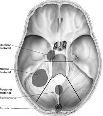
Tentorial meningioma classification
The classification system for tentorial meningiomas proposed by Gazi Yasargil is the most accurate and emphasizes the surgical anatomy.
1. T1–T2 (medial “incisural” meningioma)
2. T3–T8 (falcotentorial meningioma)
3. T4 (paramedian “intermediate” meningioma)
4. T5 (peritorcular “torcular” meningioma)
5. T6–T7 (lateral tentorial meningioma)
T1-T3 the lesions on the inner ring or lesions of the incisura - anterior, lateral and posterior.
T4 and T8 are intermediate ring lesions with T8 tumors involving the falcotentorial junction.
T5-T7 are lesions on the posterior ring, involving the torcular, transverse sinus, and transverse-sigmoid junction respectively 1).
Tentorial meningiomas are a broad and consistent category of tumors but their definition is still unclear and their classification uncertain.
Since the tentorium has a large intracranial area, tumors originating from it may vary widely in the actual location of their mass. Supratentorial, infratentorial, incisural, and posterolateral arc terms that will be used to suggest the principal location of each tumor.

