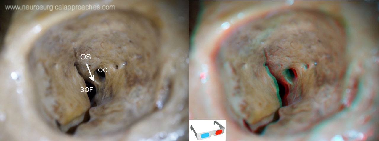
Superior orbital fissure
http://www.3dneuroanatomy.com/wp-content/uploads/2014/12/orb6.jpg
optic canal (OC)
optic strut (OS)
superior orbital fissure (SOF)
Foramen in the skull, although strictly it is more of a cleft, lying between the lesser and greater wings of the sphenoid bone.
Number 3
The medial wall of the cavernous sinus directly continues as the medial periorbita, through the superior orbital fissure.
The pericarotid sympathetic plexus coalesces to form a variable number of trunks, which exit with the sixth cranial nerve to join branches of the fifth cranial nerve entering the superior orbital fissure.
A number of important anatomical structures pass through the fissure, and these can be damaged in orbital trauma, particularly blowout fractures through the floor of the orbit into the maxillary sinus. These structures are:
superior and inferior divisions of oculomotor nerve (III) trochlear nerve (IV)
lacrimal, frontal and nasociliary branches of ophthalmic (V1). abducens nerve (VI)
superior and inferior divisions of ophthalmic vein. Inferior division also passes through the inferior orbital fissure.
sympathetic fibers from cavernous plexus
These include nonvisual sensory messages, such as pain, or motor nerves. They also serve as vascular connections.
The nerves passing through the fissure can be remembered with the mnemonic, “Live Frankly To See Absolutely No Insult” - for Lacrimal, Frontal, Trochlear, Superior Division of Oculomotor, Abducens, Nasociliary and Inferior Division of Oculomotor nerve.
It is divided into 3 parts from lateral to medial: Lateral Part transmits: lacrimal nerve, frontal nerve, trochlear nerve, meningeal branch of lacrimal artery, anastomotic branch of middle meningeal artery which anastomoses with recurrent branch of the lacrimal artery Middle Part transmits: Upper and lower divisions of the oculomotor nerve, nasociliary nerve between the two divisions of oculomotor nerve and abducent nerve Medial Part transmits: Superior ophthalmic vein and sympathetic nerves from the plexus around internal carotid artery.
The frontotemporal, so-called pterional approach has evolved with the contribution of many neurosurgeons over the past century. It has stood the test of time and has been the most commonly used transcranial approach in neurosurgery. In its current form, drilling the sphenoid wingas far down as the superior orbital fissure with or without the removal of the anterior clinoid process, thinning the orbital roof, and opening the Sylvian fissure and basal cisterns are the hallmarks of this approach.

