Retrosigmoid craniotomy
see also Retrosigmoid approach.
Position
Place the patient semiprone (45 degrees) or lateral with the falx parallel to the floor.
The incision is the length of the pinna and is located 2 cm posterior to the mastoid notch.
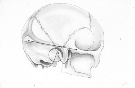
A burr hole is made at the asterion and a small craniotomy is fashioned after eggshelling edge of transverse and sigmoid sinuses.
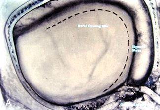
The dura is opened with triangular flaps based on the transverse and sigmoid sinuses.
The cerebellum is gently and slowly retracted medially to expose the CNs.
Water-tight dural closure is essential.
Complications
i. Facial palsy—if unable to close eyes, use natural tears q2h, Lacri-Lube, and tape eyes shut qHs
ii. Tarsorrhaphy—if after 1 year the weakness hasn't improved, perform a XII–VII anastomosis (or even after only 2 months if the nerves were directly cut)
iii. CSF leak—treat with a lumbar drain, shunt, or ENT (ear, nose, and throat) packing
iv. Vertigo—resolves with rehab
Either osteoclastic or osteoplastic bone resection can be done.
The retrosigmoid craniectomy exposes the sinus knee, the inferior border of the transverse sinus, the medial border of the sigmoid sinus and horizontal segment of the occipital squama.
The main anatomical landmarks of retrosigmoid craniotomy are transverse sinus (TS), sigmoid sinus (SS), and the confluence of both. Anatomical references and guidance based on preoperative imaging studies are less reliable in the posterior fossa than in the supratentorial region. Simple intraoperative real-time guidance methods are in demand to increase safety.
Barrero Ruiz et al. describes the localization of TS, SS, and TS-SS junction by audio blood flow detection with a micro-Doppler system.
This is an additional technique to increase safety during craniotomy and dura opening, widening the surgical corridor to secure margins without carrying risks or increasing surgical time 1).
Attention should be paid to the variable emissary veins, which lead to the sigmoid sinus. First the dura should be opened under microscopic magnification in a triangular shape in the region of the pars horizontalis to gain cerebrospinal fluid and in order to relax the cerebellum.
Then the dura has to be opened near the sinus via a curved skin incision that connects with the previous dural opening.
The lateral suboccipital retrosigmoid approach is a standard approach for removing cerebellopontine angle tumors and for microvascular decompression.
It allows enough access to the cerebellopontine angle (CPA), however the surgical view is limited to the posterior fossa and the supratentorial space is out of surgical control. Moreover, the clivus and the ventrolateral aspect of the brainstem exposure require an excessive cerebellum retraction 2).
Mastoid emissary vein is especially important from the neurosurgical point of view, because it is located in variable number in the area of the occipitomastoid suture and it can become a source of significant bleeding in surgical approaches through the mastoid process, especially in retrosigmoid craniotomy,
Retrosigmoid craniotomy


