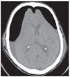
Mount Fuji sign
 Ishiwata et al. described the image produced by pneumocephalus in subdural collections that separated both frontal lobes as similar in appearance to the silhouette of the famous Fuji Volcano in Japan
1).
Mount Fuji sign is seen on cross sectional imaging and implies tension pneumocephalus is present.
The sign refers to the presence of air (pneumocephalus) between the tips of the frontal lobes giving the appearance of Mount Fuji. It suggests that the pressure of the air is at least greater than that of the surface tension of cerebrospinal fluid between the frontal lobes 2).
Appearance of this sign often warrants an immediate surgery to prevent permanent damage.
Moscovici et al. retrospectively reviewed the records of nonagenarians who had cSDH evacuation between 2006 and 2015. Postoperative computed tomographic images were evaluated for findings consistent with the MFS.
Of 45 patients, 15 patients (33%) had radiological MFS, and 3 patients (20%) with MFS required reoperation because of new blood collection. No patient required reoperation because of TP. Perioperative (30-day) mortality in patients demonstrating the MFS was 6.67% caused by cardiac arrhythmia versus 13.33% mortality in patients with no evidence of the MFS.
Mount Fuji sign in nonagenarians after cSDH evacuation is not a specific sign of TP 3).