Table of Contents
Intracranial epidermoid cyst
Epidemiology
Intracranial epidermoid cysts, are rare congenital lesions originating from the ectoderm that constitute 0.3 to 1.8 % of all intracranial neoplasms 1) 2).
The most common sites include
Cerebellopontine angle epidermoid cyst(CPA): may produce trigeminal neuralgia, especially in young patient
Suprasellar epidermoid cyst and parasellar: commonly produce bitemporal hemianopsia and optic atrophy, and only occasionally pituitary (endocrine) symptoms (including DI)
Basilar-posterior fossa specially fourth ventricle: may produce lower cranial nerve findings, cerebellar dysfunction, and/or corticospinal tract abnormalities
Sylvian fissure epidermoid cyst: may present with seizures
In the the ventricular system: occur within the 4th ventricle more commonly than any other
Approximately half of these cysts are located at the cerebellopontine angle (CPA). The most common locations of intradural epidermoids are the cerebellopontine angle cistern followed by supra- and para-sellar regions, and the fourth ventricle. Less common locations include inter-hemispheric fissure, Sylvian fissure, lateral ventricle, intracerebral, velum interpositum cistern, superior cerebellar cistern and pineal gland. They can also be extradural, usually arising in the diploic space of the calvaria, though they are less common 3).
Classification
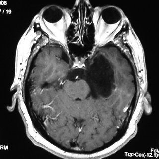
Cerebellopontine angle epidermoid cyst.
Giant intracranial epidermoid.
Pituitary infundibular epidermoid cyst
Etiology
Intracranial epidermoid cysts, also known as primary cholesteatomas are considered to arise from epithelial inclusions at the time of neural tube closure or during formation of the secondary cerebral vesicles, and have slow growth rates resembling that of the normal epidermis 4).
Pathology
Pathologically, epidermoid cysts have well-circumscribed, irregular, thin walls with squamous epithelium lining. The epithelium undergoes progressive desquamation and keratin breakdown; therefore, the cystic contents include tissue debris, keratin, water, and solid cholesterol 5).
These tumors are congenital and arise from displaced epithelial tissue between the 3rd and 5th weeks of gestation, when neural tube closure occurs.
These brain lesions have an epithelial lining that encapsulates the tumor, which consists of desquamated epidermal cell debris. Most lesions are solid, but because the desquamated cells contain cholesterol some may have a liquid cystic center. The rate of tumor growth is gradual and linear, resembling the rate of human epidermis turnover, which is unlike the exponential growth of neoplastic lesions 6).
Diagnosis
Treatment
Outcome
The most common surgical complications were transient cranial nerve palsies, occurring equally in STR and GTR cases when reported. In all postoperative epidermoid tumor cases, but particularly following STR, close follow-up with serial MRI, even years after surgery, is recommended 7).
Malignant transformation of an EC to squamous-cell carcinoma is rare; only 14 cases have been reported 8).
Meta-analysis
Shear et al., conducted a systematic review of PubMed, Web of Science, and the Cochrane Collaboration following the PRISMA guidelines. They then conducted a proportional meta-analysis to compare the pooled recurrence rates between STR and GTR in the included studies. The authors developed fixed- and mixed-effect models to estimate the pooled proportions of recurrence among patients undergoing STR or GTR. They also investigated the relationship between recurrence rate and follow-up time in the previous studies using linear regression and natural cubic spline models.
Overall, 27 studies with 691 patients met the inclusion criteria; of these, 293 (42%) underwent STR and 398 (58%) received GTR. The average recurrence rate for all procedures was 11%. The proportional meta-analysis showed that the pooled recurrence rate after STR (21%) was 7 times greater than the rate after GTR (3%). The average recurrence rate for studies with longer follow-up durations (≥ 4.4 years) (17.4%) was significantly higher than the average recurrence rate for studies with shorter follow-up durations (< 4.4 years) (5.7%). The cutoff point of 4.4 years was selected based on the significant relationship between the recurrence rate of both STR and GTR and follow-up durations in the included studies (p = 0.008).
STR is associated with a significantly higher rate of epidermoid tumor recurrence compared to GTR. Attempts at GTR should be made during the initial surgery with efforts to optimize success. Surgical expertise, as well as the use of adjuncts, such as intraoperative MRI and neuromonitoring, may increase the likelihood of completing a safe GTR and decreasing the long-term risk of recurrence. The most common surgical complications were transient cranial nerve palsies, occurring equally in STR and GTR cases when reported. In all postoperative epidermoid tumor cases, but particularly following STR, close follow-up with serial MRI, even years after surgery, is recommended 9).
Case series
Vaz-Guimaraes et al., from Pittsburgh, Houston, and Toronto retrospectively reviewed the medical records of 21 patients who underwent endoscopic endonasal surgery for epidermoid and dermoid cyst resection at the University of Pittsburgh Medical Center between January 2005 and June 2014. Surgical outcomes and variables that might affect the extent of resection and complications were analyzed.
Total resection (total removal of cyst contents and capsule) was achieved in 8 patients (38.1%), near-total resection (total removal of cyst contents, incomplete removal of cyst capsule) in 9 patients (42.9%), and subtotal resection (incomplete removal of cyst contents and capsule) in 4 patients (19%). Larger cyst volume (≥ 3 cm3) and intradural location (15 cysts) were significantly associated with nontotal resection (p = 0.008 and 0.0005, respectively). In the whole series, surgical complications were seen in 6 patients (28.6%). No complications were observed in patients with extradural cysts. Among the 15 patients with intradural cysts, the most common surgical complication was postoperative Cerebrospinal fluid fistula (5 patients, 33.3%), followed by postoperative intracranial infection (4 patients, 26.7%). Larger cysts and postoperative Cerebrospinal fluid fistula were associated with intracranial infection (p = 0.012 and 0.028, respectively). Subtotal resection was marginally associated with intracranial infection when compared with total resection (p = 0.091). All patients with neurological symptoms improved postoperatively with the exception of 1 patient with unchanged abducens nerve palsy.
Endoscopic endonasal approaches may be effectively used for resection of epidermoid and dermoid cysts in carefully selected cases. These approaches are recommended for cases in which a total or near-total resection is possible in addition to a multilayer cranial base reconstruction with vascularized tissue to minimize the risk of intracranial infection 10).
