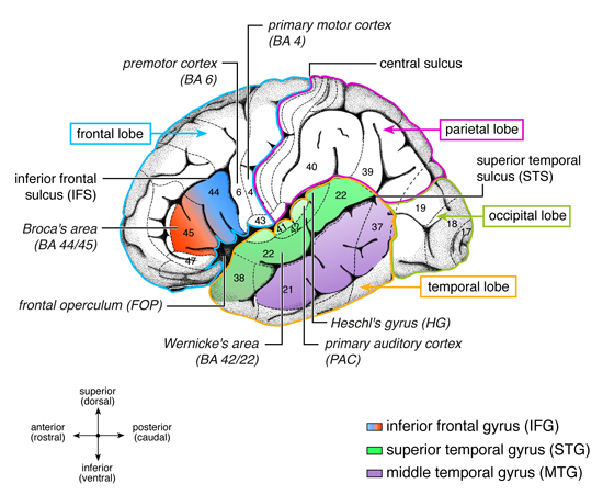
Heschl's Gyrus
Richard Ladislaus Heschl
In 1849 he received his medical doctorate from the University of Vienna, where in 1850 he became a “first assistant” to Carl von Rokitansky (1804-1878). In 1854 he was appointed professor of anatomy at the medical-surgical school in Olomouc, and during the following year became a professor of pathological anatomy in Krakow. In 1861 he became a professor at the medical-surgical school at Graz (from 1863 a full professor), serving as university rector in 1864-65. In 1875 he returned to the University of Vienna. After his death, his position at Vienna was filled by Hans Kundrat (1845-1893).
Heschl is credited as the first physician to describe the transverse temporal gyrus or “Heschl's gyrus”, located in the primary auditory cortex. This anatomical structure processes incoming auditory information.
That portion of the superior temporal gyrus that lies inside the banks of the lateral fissure (the temporal operculum). This area is also referred to as the transverse gyri of Heschl as the individual gyri are oriented in a lateral–medial plane within the operculum (as opposed to a more anterior–posterior or inferior–superior plane of most of the cortical gyri).
This region is the primary cortical projection area for auditory information coming from the medial geniculates, the specific auditory relay nuclei of the thalamus. Because of the extensive bilateral projections that characterize the auditory system, unilateral lesions restricted to Heschl’s gyrus would be expected to produce only very subtle deficits and may be limited to increased difficulty with sound localization. While quite rare, bilateral lesions to this structure could result in deafness.
Local field potentials were recorded from 8 patients implanted with depth-electrodes in Heschl's gyrus and the planum temporale (55 recording sites in total), usually considered as human primary and secondary auditory cortices. Using a frequency-tagging approach, we show that both low-frequency (<30 Hz) and high-frequency (>30 Hz) neural activities in these structures faithfully track auditory rhythms through frequency-locking to the rhythm envelope. A selective gain in amplitude of the response frequency-locked to the beat frequency was observed for the low-frequency activities but not for the high-frequency activities, and was sharper in the planum temporale, especially for the more challenging syncopated rhythm. Hence, this gain process is not systematic in all activities produced in these areas and depends on the complexity of the rhythmic input. Moreover, this gain was disrupted when the rhythm was presented at fast speed, revealing low-pass response properties which could account for the propensity to perceive a beat only within the musical tempo range. Together, these observations show that, even though part of these neural transforms of rhythms could already take place in subcortical auditory processes, the earliest auditory cortical processes shape the neural representation of rhythmic inputs in favor of the emergence of a periodic beat 1).
