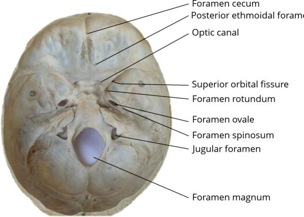
Foramen spinosum
 One of two foramina located in the base of the human skull, on the sphenoid bone. It is situated just anterior to the spine of the sphenoid bone, and just lateral to the foramen ovale. The middle meningeal artery, middle meningeal vein, and the meningeal branch of the mandibular nerve pass through the foramen.
The foramen spinosum is often used as a landmark in neurosurgery, due to its close relations with other cranial foramen. It was first described by Jakob Benignus Winslow in the 18th century.
The foramen spinosum is a foramen through the sphenoid bone situated in the middle cranial fossa.
It is one of two foramina in the greater wing of the sphenoid bone. The foramen ovale is one of these two cranial foramina, situated directly anterior and medial to the foramen spinosum.[
The spine of sphenoid falls medial and posterior to the foramen. Lateral to the foramen is the mandibular fossa, and posterior is the Eustachian tube.
The foramen spinosum varies in size and location. The foramen is rarely absent, usually unilaterally, in which case the middle meningeal artery enters the cranial cavity through the foramen ovale.
It may be incomplete, which may occur in almost half of the population. Conversely, in a minority of cases (less than 1%), it may also be duplicated, particularly when the middle meningeal artery is also duplicated.
The foramen may pass through the sphenoid bone at the apex of the spinous process, or along its medial surface.