Diffusion-weighted magnetic resonance imaging indications
Diffusion-weighted magnetic resonance imaging (DWI) is a specialized MRI technique that measures the random movement (diffusion) of water molecules in tissue. It is increasingly used in the diagnosis of glioblastoma recurrence due to its ability to provide insights into tissue cellularity and the integrity of cell membranes, which change significantly in tumor recurrence.
Results did not show usefulness of the Diffusion-weighted magnetic resonance imaging and T1-weighted images for assessing the consistency of pituitary macroadenomas, nor as a predictor of the degree of surgical resection 1)
It is considered useful not only for the detection of acute ischemic stroke but also for the characterization and differentiation of brain tumors and brain abscess.
There are numerous applications of DWI/DTI in brain tumors: (1) determination of grade and histologic subtype, (2) evaluation of peritumoral edema and assessment of pathways of tumor infiltration, (3) quantitative measurement and monitoring of the response to therapy, and (4) discrimination between necrosis and tumor recurrence (eg, radiation and chemotherapy).
Differences in diffusion properties of low- and high-grade tumors are caused by several factors including different tumor cellularity and nucleus to cytoplasm ratio. In contrast to high-grade tumors, low-grade tumors are characterized by hypocellularity, low nucleus to cytoplasm ratio, and large extracellular spaces, which is typically represented by high MD/ADC values 2) 3).
Studies involving coronary artery bypass graft surgery, carotid endarterectomy, or interventional surgery have demonstrated new small ischemic brain lesions using DWI.
Normally water protons have the ability to diffuse extracellularly and loose signal. High intensity on DWI indicates restriction of the ability of water protons to diffuse extracellularly. Restricted diffusion is seen in abscesses, epidermoid cysts and acute infarction (due to cytotoxic edema).
Some tumors: most tumors are dark on DWI, but highly cellular tumors may have decreased diffusion (bright on DWI) (e.g. epidermoids, some meningiomas…)
In cerebral abscesses the diffusion is probably restricted due to the viscosity of pus, resulting in a high signal on DWI.
In most tumors there is no restricted diffusion - even in necrotic or cystic components. This results in a normal, low signal on DWI.
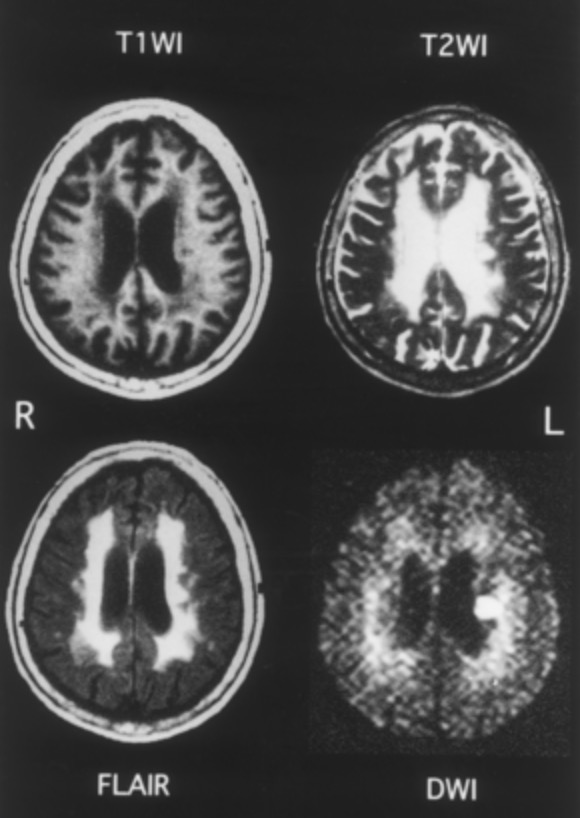
MRI of 72-year-old woman admitted because of right hemiparesis. MRI was performed 7 days after onset. T1-weighted imaging revealed multiple low-intensity areas around the ventricle, and both T2-weighted imaging and FLAIR showed an area of severe periventricular hyperintensity with suspected multiple high-intensity lesions. DWI showed a high-intensity area that coincided with clinical features on the left corona radiata.
DWI or MRA conducted immediately after Aneurysm clipping may be affected by artifacts resulting from the surgical procedure, such as intracranial air or motion artifacts from the patient.
DWI was performed using two-dimensional, single-shot, spin-echo, echo planar imaging of the entire brain with the following parameters: echo time (TE), 50; repetition time (TR), infinite; B, 1000 s/mm2; field of view (FOV), 24 × 24 cm; flip angle, 90°; imaging matrix, 128 × 128; slice thickness, 5.5 mm with a 1.5-mm gap; and number of slices, 20. Three- dimensional T1 fast field echo time-of-flight MRA of the circle of Willis was performed using the following parameters: flip angle, 18°; TR, 25 ms; TE, 3.5 ms; slice thickness, 1.2 mm; FOV, 20 × 20; matrix size, 512 × 205; number of slices, 132– 160; slice gap, 0.6 mm.
Any new hyperintensities observed using postoperative DWI were interpreted as new ischemic lesions that developed after aneurysm clipping 4).
