Broca's area
A region of the frontal lobe that has been named after Paul Broca.
Broca’s Area is involved with articulated language.
Language dysfunction is a common presentation for patients with a glioma that involves language areas. see Broca's aphasia.
An easy way to delimitate Broca's area on the dominant hemisphere is by localizing four craniometrical points
a) the Stephanion, b) 2 cm posterior to the Stephanion C) the anterior Sylvian point and D) the inferior rolandic point (IRP).
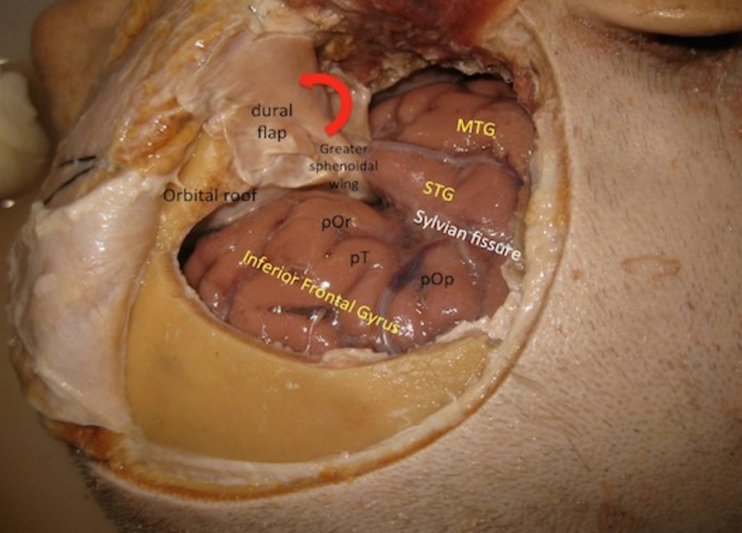
The bone flap has been removed and the dura mater has been opened as a flap pediculated towards the greater sphenoid wing previously roungered to improve parasellar visualization. Sylvian fissure, Inferior frontal gyrus, Superior temporal gyrus and Middle temporal gyrus are exposed. Three pars of parasylvian inferior frontal gyrus must be distinguished: pars orbitalis (pOr) in relation to the orbital roof; pars triangularis (pT) the widest area of sylvian fissure (good place for start opening of sylvian fissure); pars opercularis (pOp) where Broca’s Area is located.
Or the Broca area /broʊˈkɑː/ or /ˈbroʊkə/ is a region in the frontal lobe of one hemisphere (usually the left) of the brain with functions linked to speech production.
The production of language has been linked to the Broca's area since Paul Broca reported impairments in two patients.
They had lost the ability to speak after injury to the posterior inferior frontal gyrus of the brain.
Since then, the approximate region he identified has become known as Broca's area, and the deficit in language production as Broca's aphasia.
Broca's area is now typically defined in terms of the pars opercularis and pars triangularis of the inferior frontal gyrus, represented in Brodmann's cytoarchitectonic map as areas 44 and 45 of the dominant hemisphere.
Studies of chronic aphasia have implicated an essential role of Broca's area in various speech and language functions. Further, functional MRI studies have also identified activation patterns in Broca's area associated with various language tasks. However, slow destruction of the Broca's area by brain tumors can leave speech relatively intact suggesting its functions can shift to nearby areas in the brain.
Corns et al. describe the case of a patient with recurrent glioblastoma encroaching on Broca's area (eloquent brain). Gross total resection of the tumour was achieved by combining two techniques, awake craniotomy to prevent damage to eloquent brain and 5 aminolevulinic acid fluorescence guided resection to maximise the extent of tumour resection.This technique led to gross total resection of all T1-enhancing tumour with the avoidance of neurological deficit. They recommend this technique in patients when awake surgery can be tolerated and gross total resection is the aim of surgery 1).
