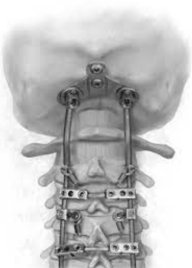
In a retrospective cohort study Shahin et al. from the Doernbecher Children’s Hospital and Oregon Health & Science University, Portland published in the Journal of Neurosurgery Pediatrics to assess whether screw‑fixed autologous rib grafts improve fusion rates in pediatric occipitocervical fusion (OCF), and validate a novel imaging-based fusion grading scale independent of graft type. Screw‑anchored rib autograft achieved 100 % solid fusion at ≥3 months (n=16), compared to 57 % fusion (4/7) and 43 % resorption/pseudarthrosis in standard allograft/BMP group (p=0.0066). The new 0–2 radiographic grade correlated well with CT-defined outcomes 11)
Critical review
1. Study design & cohort: Retrospective, single‑institution, relatively small sample (n=21 total; final rib‑graft cohort n=17 minus one without CT). Comparison spans two eras (2015–2016 vs. 2016–2022), risks secular trends or surgeon learning‑curve bias.
2. Intervention vs. control: Cohort 1 received standard instrumentation with allograft/BMP; cohort 2 received screw‑fixed rib graft. But several cohort 2 cases were revisions from cohort 1, confounding the groups. No randomization.
3. Outcomes & follow-up: Fusion assessed at ≥3 months by blinded neuroradiologists with a 0–2 grading scale—clear and reproducible. However, mid / long‑term (>1 year) follow-up beyond early fusion rate not well characterized.
4. Results interpretation: Dramatic fusion improvement is compelling, but may reflect both graft technique and instrumentation changes over time. Lack of halo/BMP/lab comparisons limiting.
5. Radiographic grading scale: Solid concept, but needs external validation across graft types and institutions.
6. Safety & complications: No donor‑site morbidity or hardware failures reported over 5+ years. But small sample limits detection of rare complications.
7. Limitations:
