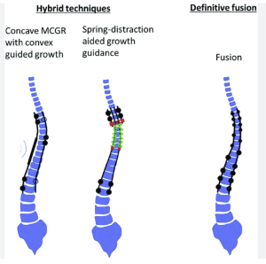
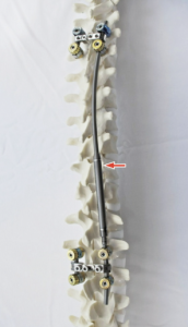
All‑pedicle screw posterior spinal fusion after magnetically controlled growing rods in walking early‑onset scoliosis patients: radiographic and surgical outcomes

– Strengths:

  1. Focus on a clinically relevant patient group (walking EOS post-MCGR).
  2. Objective measurement of both coronal and sagittal parameters with adequate follow-up.

– Limitations:

  1. Small sample (n=27) limits statistical power and heterogeneity evaluation.
    Read more

Failure to replace removed growth friendly implants results in deteriorating radiographic outcomes

In a registry-based comparative cohort study (early onset scoliosis patients after implant removal) Matan S Malka et al. from the Morgan Stanley Children’s Hospital (Columbia Univ, New York). Arkansas Children’s Hospital; Shriners Philadelphia; Seattle Children’s Hosp. published in Spine Deformity Journal, to evaluate if re-implanting growth-friendly constructs within 12 months after implant removal (ROI) stabilizes deformity compared to observation-only. Early re-implantation (< 12 mo post-ROI) significantly reduces 2‑year coronal Cobb progression compared to no replacement 1).

Critical Review

– Strengths:

Multicenter registry with well-defined exposure groups.

Radiographic outcomes measured at a meaningful 2‑year follow-up.

Statistically robust with p-values: Cobb 81° vs 53° (p=0.003); progression ≥5°: 64% vs 30% (p=0.04)

– Limitations:

Read more

Autologous rib graft augmentation for occipitocervical fusion in pediatric patients and a novel radiographic grading scale

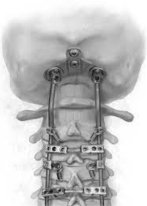
In a retrospective cohort study Shahin et al. from the Doernbecher Children’s Hospital and Oregon Health & Science University, Portland published in the Journal of Neurosurgery Pediatrics to assess whether screw‑fixed autologous rib grafts improve fusion rates in pediatric occipitocervical fusion (OCF), and validate a novel imaging-based fusion grading scale independent of graft type. Screw‑anchored rib autograft achieved 100 % solid fusion at ≥3 months (n=16), compared to 57 % fusion (4/7) and 43 % resorption/pseudarthrosis in standard allograft/BMP group (p=0.0066). The new 0–2 radiographic grade correlated well with CT-defined outcomes 11)

1. Study design & cohort: Retrospective, single‑institution, relatively small sample (n=21 total; final rib‑graft cohort n=17 minus one without CT). Comparison spans two eras (2015–2016 vs. 2016–2022), risks secular trends or surgeon learning‑curve bias.

2. Intervention vs. control: Cohort 1 received standard instrumentation with allograft/BMP; cohort 2 received screw‑fixed rib graft. But several cohort 2 cases were revisions from cohort 1, confounding the groups. No randomization.

3. Outcomes & follow-up: Fusion assessed at ≥3 months by blinded neuroradiologists with a 0–2 grading scale—clear and reproducible. However, mid / long‑term (>1 year) follow-up beyond early fusion rate not well characterized.

4. Results interpretation: Dramatic fusion improvement is compelling, but may reflect both graft technique and instrumentation changes over time. Lack of halo/BMP/lab comparisons limiting.

5. Radiographic grading scale: Solid concept, but needs external validation across graft types and institutions.

6. Safety & complications: No donor‑site morbidity or hardware failures reported over 5+ years. But small sample limits detection of rare complications.

7. Limitations:

Read more