Novel planning pipeline utilizing the Surgical Theater system for pediatric epilepsy surgery

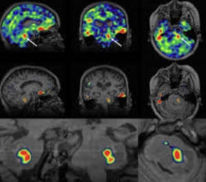
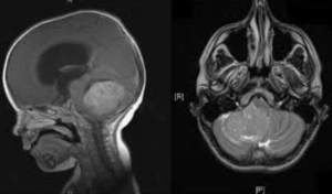
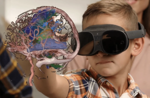
In a retrospective institutional experience with illustrative cases Shields LB et al. from the Norton Neuroscience Institute, Louisville published in Epilepsia Open to evaluate the role of the Surgical Theater (ST) 3D visualization system in enhancing presurgical planning for pediatric epilepsy surgery. The ST system enabled integration of multimodal imaging into immersive 3D models, improving collaborative surgical planning, enhancing intraoperative navigation, and allowing VR-based procedural rehearsal. It demonstrated utility across 85 cases and is posited as a promising adjunct for pediatric epilepsy surgical workflows 1)

This is a descriptive, non-comparative experience report centered on implementing the Surgical Theater (ST) system in a pediatric epilepsy context. While the authors present a visually compelling and potentially transformative workflow for presurgical planning, the study is methodologically weak—it lacks controls, quantifiable outcomes, or statistical rigor. The “results” are largely anecdotal, with 4 case examples insufficiently discussed in terms of surgical impact or clinical outcomes.

Read more

All‑pedicle screw posterior spinal fusion after magnetically controlled growing rods in walking early‑onset scoliosis patients: radiographic and surgical outcomes

– Strengths:

  1. Focus on a clinically relevant patient group (walking EOS post-MCGR).
  2. Objective measurement of both coronal and sagittal parameters with adequate follow-up.

– Limitations:

  1. Small sample (n=27) limits statistical power and heterogeneity evaluation.

    Read more

Failure to replace removed growth friendly implants results in deteriorating radiographic outcomes

In a registry-based comparative cohort study (early onset scoliosis patients after implant removal) Matan S Malka et al. from the Morgan Stanley Children’s Hospital (Columbia Univ, New York). Arkansas Children’s Hospital; Shriners Philadelphia; Seattle Children’s Hosp. published in Spine Deformity Journal, to evaluate if re-implanting growth-friendly constructs within 12 months after implant removal (ROI) stabilizes deformity compared to observation-only. Early re-implantation (< 12 mo post-ROI) significantly reduces 2‑year coronal Cobb progression compared to no replacement 1).

Critical Review

– Strengths:

Multicenter registry with well-defined exposure groups.

Radiographic outcomes measured at a meaningful 2‑year follow-up.

Statistically robust with p-values: Cobb 81° vs 53° (p=0.003); progression ≥5°: 64% vs 30% (p=0.04)

– Limitations:

Read more

Comparative assessment of stereoelectroencephalography and subdural electrodes in invasive epilepsy monitoring: a systematic review and meta‑analysis

In a systematic review and metaanalysis of double‑arm comparative studies Bandopadhay et al. from the Houston Methodist Hospital published in the Journal of Neurosurgery to compare safety and seizure‑outcome profiles of stereoelectroencephalography (SEEG) vs. subdural electrodes (SDE) in pharmacoresistant epilepsy using quantitative double‑arm data SEEG demonstrated a higher rate of favorable seizure outcomes (RR 1.14, 95% CI 1.02–1.27; p=0.02) and lower complication rates overall (RR 0.49, 95% CI 0.37–0.66; p<0.00001). The benefit was significant in general adult cohorts but less pronounced in pediatric or older groups 1).


Strengths:

  • Restricting to double‑arm designs reduces cross‑study heterogeneity.
  • Large pooled cohort: 1,632 SEEG vs. 1,482 SDE patients.
  • Age‑stratified subgroup analysis adds nuance to applicability.

Limitations:

  • Potential for publication bias—likely underreporting of negative or null comparative studies.

    Read more

Management of patients discharged from the hospital after VPS surgery

In a Letter to the Editor Lu et al. published in the Journal of Neurosurgery to discuss ventriculoperitoneal shunt management strategies for discharged patients 1).

Critical Appraisal

– Strengths:

  1. Highlights a clinically important gap—post-discharge VPS care.
  2. Sparks important discussion on outpatient monitoring and follow-up protocols.

– Limitations:

  1. Absence of abstract/data: no study design, patient numbers, follow-up length or outcomes described.

    Read more

Autologous rib graft augmentation for occipitocervical fusion in pediatric patients and a novel radiographic grading scale

In a retrospective cohort study Shahin et al. from the Doernbecher Children’s Hospital and Oregon Health & Science University, Portland published in the Journal of Neurosurgery Pediatrics to assess whether screw‑fixed autologous rib grafts improve fusion rates in pediatric occipitocervical fusion (OCF), and validate a novel imaging-based fusion grading scale independent of graft type. Screw‑anchored rib autograft achieved 100 % solid fusion at ≥3 months (n=16), compared to 57 % fusion (4/7) and 43 % resorption/pseudarthrosis in standard allograft/BMP group (p=0.0066). The new 0–2 radiographic grade correlated well with CT-defined outcomes 11)

1. Study design & cohort: Retrospective, single‑institution, relatively small sample (n=21 total; final rib‑graft cohort n=17 minus one without CT). Comparison spans two eras (2015–2016 vs. 2016–2022), risks secular trends or surgeon learning‑curve bias.

2. Intervention vs. control: Cohort 1 received standard instrumentation with allograft/BMP; cohort 2 received screw‑fixed rib graft. But several cohort 2 cases were revisions from cohort 1, confounding the groups. No randomization.

3. Outcomes & follow-up: Fusion assessed at ≥3 months by blinded neuroradiologists with a 0–2 grading scale—clear and reproducible. However, mid / long‑term (>1 year) follow-up beyond early fusion rate not well characterized.

4. Results interpretation: Dramatic fusion improvement is compelling, but may reflect both graft technique and instrumentation changes over time. Lack of halo/BMP/lab comparisons limiting.

5. Radiographic grading scale: Solid concept, but needs external validation across graft types and institutions.

6. Safety & complications: No donor‑site morbidity or hardware failures reported over 5+ years. But small sample limits detection of rare complications.

7. Limitations:

Read more

Sodium MRI in Pediatric Brain Tumors

In a narrative review Bhatia et al. from the Children’s Hospital of Philadelphia, Radiological Sciences Laboratory, School of Medicine, Stanford University, published in the American Journal of Neuroradiology to explore the potential of sodium-23 MRI (^23Na-MRI) as a noninvasive imaging modality to assess physiological and biochemical changes in pediatric brain tumors and concluded that is a promising, noninvasive imaging modality capable of providing unique physiological and biochemical information that is not accessible through conventional MRI techniques

This narrative review attempts to position ^23Na-MRI as a frontier imaging technique for pediatric brain tumors. It lauds the modality’s potential to reveal sodium-dependent physiological alterations — but quickly devolves into technological evangelism with minimal clinical anchoring. The piece is high on optimism, low on pragmatism, and entirely devoid of data-supported clinical outcomes.

The article enthusiastically describes the theoretical virtues of sodium MRI — sensitivity to cell integrity, ionic gradients, extracellular space — but offers no compelling clinical cases, no comparative metrics, and no outcome data. What remains is a speculative wish list, presented as a roadmap. The authors confuse imaging potential with diagnostic utility, a common pitfall in radiology reviews driven by physics rather than patient care.

“Exciting” is not a scientific category.

Read more

Exploring IDH1 and IDH2 Mutations in Paediatric Medulloblastoma

In a retrospective molecular analysis of 23 pediatric medulloblastoma samples, Motaz Fadul et al. from – King Abdulaziz University, Jeddah – King Faisal Specialist Hospital and Research Center, Jeddah – Umm Al-Qura University, Makkah – King Saud University, Riyadh – King Abdulaziz Medical City, Jeddah – King Fahad General Hospital, Jeddah Published in: Folia Biologica (Praha), 2025;71(2):73-78

This study sought to:

Findings:

  • ❌ No IDH1 or IDH2 mutations were detected in any of the 23 tumor samples.
  • 🧬 β-Catenin expression showed no significant correlation with PFS.
  • 📉 Median PFS was 383 days, suggesting modest clinical outcomes.

These findings reinforce the understanding that IDH mutations, prevalent in adult gliomas, are not typical of paediatric medulloblastomas, possibly due to divergent tumorigenic pathways and distinct cellular origins 2)


This study contributes marginally to the existing molecular profiling literature on paediatric medulloblastoma:

  • ✅ Reaffirms known absence of IDH mutations
  • ❗ Sample size (n=23) limits statistical power
  • ❗ Overreliance on β-catenin expression, without subgroup stratification (e.g. WNT/SHH)
  • ❗ Methodology (PCR + Sanger sequencing) lacks genomic depth in the era of next-generation sequencing
  • ❌ No novel biomarkers or clinical correlates were identified

    Read more