LINC01783 Promotes Glioma Tumorigenesis by Enhancing GATA3 Expression Through CBP-Mediated H3K27 Acetylation to Suppress PTEN Expression

LINC01783

Type of Study: In vitro and in vivo molecular mechanistic investigation First Author: Shaocai Hao et al. Author Affiliations:

  • Department of Neurosurgery, General Hospital of Ningxia Medical University, Yinchuan, Ningxia, China

Journal: Biofactors DOI: 10.1002/biof.70029 PMID: 40546096 Publication Date: May–June 2025 Title: LINC01783 Promotes Glioma Tumorigenesis by Enhancing GATA3 Expression Through CBP-Mediated H3K27 Acetylation to Suppress PTEN Expression

To elucidate the oncogenic function of the long intergenic non-coding RNA LINC01783 in glioma progression, focusing on its effect on GATA3 expression and PTEN suppression via CBP-mediated H3K27 acetylation.

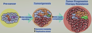
LINC01783 is significantly upregulated in glioma tissues and enhances glioma progression by promoting GATA3 expression through CBP-mediated H3K27 acetylation. This, in turn, transcriptionally represses PTEN, contributing to increased tumor cell proliferation and stemness.

  • Sample opacity: No clear details on glioma sample number, subtype stratification, or clinical metadata; undermines reproducibility and clinical significance.
  • In vivo data insufficiently controlled: No information on animal randomization, group sizes, or blinding procedures. Xenograft conclusions are weakly supported.
  • Epigenetic mechanistic oversimplification: Attribution of GATA3 regulation solely to CBP-H3K27ac is unconvincing; alternative pathways and compensatory mechanisms are unexamined.
  • Lack of causal proof: The PTEN axis is emphasized, but whether GATA3 mediates all observed phenotypes is not demonstrated.
  • No translational bridge: No therapeutic agent, inhibitor, or antisense strategy explored. The leap to “potential therapeutic target” is scientifically unfounded.
    Read more

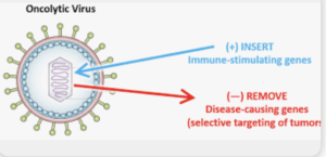
Oncolytic virus‑mediated immunomodulation in glioblastoma: Insights from clinical trials and challenges

In a Review Raziye Piranlioglu *et al.* from

Affiliations Harvey Cushing Neuro‑oncology Laboratories, Dept. Neurosurgery, Brigham and Women’s Hospital, Boston, MA, USA; Dana‑Farber Cancer Institute, Boston, MA, USA

published in *Seminars in Immunology* with the Purpose to synthesize data from clinical trials of oncolytic viruses (OVs) in glioblastoma, evaluating immunomodulatory effects, delivery strategies, and challenges in assessing immune responses. They concluded that Oncolytic virus therapy is well tolerated in GBM trials and can convert the immunosuppressive microenvironment into an immunologically active state. However, limitations in post‑treatment sampling and delivery methods impede full understanding of biological mechanisms.


This review is a rehash of well‑known take‑home messages, offering little in the way of novel synthesis or incisive critique. The authors lean heavily on canonical trials (e.g., oHSV, adenovirus) but fail to integrate preclinical correlates from myeloid-targeting strategies, such as macrophage polarization dynamics or MDSC modulation. There’s no fresh mechanism, no meta‑analysis of response rates, and no exploration of why most trials remain phase I with limited impact. Sample‑scarcity is once again highlighted as a blocker—but no alternative trial designs (e.g., neoadjuvant window cohorts, liquid biopsy) are proposed. In short, the review scratches the surface of challenges without pushing the field forward.

Read more

A national study of neurosurgical residency competency development

In a retrospective observational cohort study Using national milestone data from 2478 neurosurgery residents across 120 U.S. programs (2018–2022), with descriptive statistical analysis Khalid et al.evaluate the progression of neurosurgical residents across the 6 ACGME core competencies and 20 subcompetencies, specifically: Assessing how many residents reach level 4 proficiency by the final year (PGY-7). Identifying patterns of co-occurring deficiencies in competencies. They conclude that neurosurgery residents demonstrate substantial milestone progression throughout training, but gaps remain—particularly in specialized clinical skills and self-assessment (Reflective Practice). Nearly 45% fail to reach level 4 in at least one subcompetency by PGY-7. These deficiencies are concentrated in areas often covered during fellowship training (e.g., epilepsy, pain, peripheral nerve). Therefore, residency programs may need to enhance exposure to these areas or redefine competency expectations. The authors recommend: Targeted educational interventions

Specialized procedural training To ensure that all residents achieve the necessary competencies for independent practice 1)


This study mistakes numerical progression in a checklist for actual neurosurgical maturity. “Milestones” are treated as objective truths, when in reality they are administrative fictions imposed top-down by ACGME to simulate accountability. The implicit assumption—that every resident must hit an arbitrary “level 4” to be considered competent—is never questioned. The authors do not interrogate what level 4 means, who defines it, or whether it maps to meaningful clinical outcomes. Instead, they deliver descriptive statistics masquerading as insights.

Read more

Post-traumatic hydrocephalus after decompressive craniectomy: a multidimensional analysis of clinical, radiological, and surgical risk factors

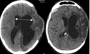
In a retrospective observational cohort study Romualdo et al. from the Department of Neurosurgery Faculty of Medicine, Technische Universität Dresden University Hospital Carl Gustav Carus published in the Neurosurgical Review to identify clinical, radiological, and surgical risk factors associated with the development of shunt-dependent posttraumatic hydrocephalus (PTH) in patients who underwent decompressive craniectomy following severe traumatic brain injury (TBI). Shunt-dependent post-traumatic hydrocephalus (PTH) occurred in 27% of patients after decompressive craniectomy for severe TBI. Independent risk factors included older age, basal cistern subarachnoid hemorrhage, post-traumatic ischemic infarcts, transcalvarial herniation, subdural hygroma, and progressive contusion hemorrhages. Surgical parameters were not predictive. Patients requiring shunt placement had significantly worse neurological outcomes 5).


🚨 The Illusion of Multidimensionality Despite claiming a “multidimensional” analysis, the study delivers a monotonous list of obvious associations—many of which have been reported in the literature for over a decade. Subarachnoid hemorrhage, infarction, hygroma, contusion progression… yes, thank you, we knew that. What’s new? Almost nothing.

Read more

Outcomes of CT-Guided Targeted Epidural Patching for Lateral Dural Tears in Spontaneous Intracranial Hypotension: A Multicenter Retrospective Cohort Study

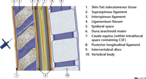
In a multicenter retrospective cohort study, Callen et al. — from the University of Colorado Anschutz, Kaiser Permanente Santa Clara, University of Freiburg, Cambridge University Hospitals, Newcastle upon Tyne Hospitals, Guy’s & St Thomas’s / King’s College Hospitals, and King’s College London — published in the American Journal of Neuroradiology, the clinical and radiologic outcomes of CT-guided epidural patching in patients with lateral dural tear causing spontaneous intracranial hypotension (SIH). The study also aimed to determine whether anatomic factors (e.g., herniated arachnoid pouch) or procedural variables (e.g., patch volume, material, approach) predict treatment success.

CT-guided patching led to complete symptom resolution in approximately one-third of patients. The presence of a herniated arachnoid pouch was associated with lower radiologic resolution of CSF collections. Procedural variables — such as patch type, approach, and volume — were not associated with outcomes. Notably, some patients experienced clinical improvement despite persistent CSF collections, highlighting the need for long-term follow-up and cautious reliance on imaging alone.

1)

Read more

Long-Term Mortality of Patients With Head Injuries—A 10-Year Follow-up Study With Population Controls

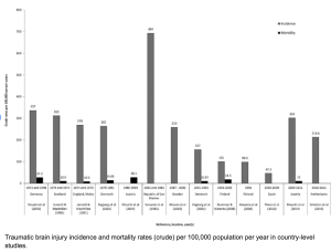
In a retrospective, population-based cohort study with matched controls and longitudinal follow-up, Heinonen et al. from Tampere University Hospital, Helsinki University Hospital, and Harvard Medical School in the Neurosurgery Journal compared 10-year survival rates and causes of death between patients with traumatic head injuries treated at a university hospital and matched population controls. They aimed to identify factors associated with long-term mortality after TBI.

Patients with head injuries exhibited significantly reduced long-term survival compared to matched controls, even after excluding early mortality. However, patient-related characteristics (e.g., comorbidities, lifestyle factors) — more than injury severity itself — appeared to drive this increased mortality risk.

Notably, even patients without documented TBI (likely mild or undiagnosed) showed decreased survival, suggesting an under-recognized long-term impact of head injury across all severity levels 2).


In this population-based cohort study, the authors track 10-year mortality in over 1,900 patients with head injuries versus 9,600 matched controls. Unsurprisingly, trauma patients die more — especially from alcohol, accidents, and “patient characteristics.” The conclusion? It’s not the injury; it’s the person. This study doesn’t just underdeliver — it underthinks.

The study’s main conclusion — that patient-related factors, not injury severity, explain increased mortality — is not only reductive but evasive. The term “patient characteristics” serves as a statistical landfill for all the unmeasured, uncontrolled, and misunderstood variables: mental health, addiction, social deprivation, neurobehavioral sequelae… all dumped under one lazy label.

Rather than confront the neuropsychiatric aftermath of head trauma, the authors retreat behind correlational shields.

❝They died because of who they were, not what happened to them.❞ — That’s not science. That’s resignation.

Read more

Maximizing Tumor Resection and Managing Cognitive Attentional Outcomes: Measures of Impact of Awake Surgery in Glioma Treatment

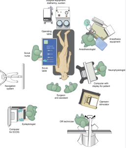
In a retrospective observational study Zigiotto et al. from the S. Chiara University-Hospital, Azienda Provinciale per i Servizi Sanitari, Trento, published in the Neurosurgery Journal on 64 glioma patients who underwent awake surgery (AwS) or asleep surgery (AsS), with neuropsychological and imaging follow-up. They evaluated the impact of awake surgery on attentional outcomes in glioma patients, and analyzed whether greater extent of tumor resection correlates with transient cognitive (attentional) decline, especially in relation to lesions within the default mode network. Awake surgery allows for more extensive supramaximal resection and is associated with longer overall survival, particularly in patients with glioblastomas. However, it also leads to a higher rate of transient postoperative attentional dysfunction, likely due to resection in attention-related brain networks. The study suggests that patient selection and intraoperative cognitive monitoring should be optimized in future glioma surgery 5).


This retrospective study compares awake versus asleep craniotomy in 64 glioma patients, using simple attention tests before and after surgery. The authors claim that awake craniotomy (AwC) allows more extensive tumor resection and leads to longer survival, albeit at the cost of transient attentional dysfunction.

The title promises a nuanced exploration of cognitive outcomes. What it delivers is a reduction of “attention” to the Trail Making Test Part A and a visual search task — an embarrassingly narrow lens for such a multidimensional construct. The study purports to evaluate the impact of surgery on attention, yet fails to define attention, stratify its subtypes, or provide any neuropsychological depth. This is not a cognitive study — it’s a surgical paper pretending to be one.

Read more

Reentry Technique for Rescue Recanalization of Carotid Near-Total Occlusion after Subintimal Penetration

In a case report, Tran et al., from the Department of Neurosurgery, University Medical Center Ho Chi Minh City; the Vascular Department, Cho Ray Hospital, Ho Chi Minh City, Vietnam; and the Department of Neurosurgery, Taichung Veterans General Hospital and National Chung Hsing University, Taichung, Taiwan, published in the American journal of neuroradiology that in selected patients with near-total carotid artery occlusion, where subintimal dissection occurs during endovascular access, the use of a rescue reentry technique with a reentry catheter can be a safe and effective method for achieving successful recanalization and restoring luminal patency 2).


The authors present a single-patient case in which a reentry catheter was used to regain access to the true lumen after unintentional subintimal dissection during an attempt to stent a near-occluded carotid artery. Technical success was achieved, and the patient recovered well.

This article epitomizes a growing trend in low-yield case reports: elevate the ordinary by inflating the vocabulary. A standard bailout technique, long adapted from peripheral interventions, is glorified as a “rescue reentry technique” as if it were a novel paradigm. This is not a new technique — it’s complication management.

🧩 What they call “rescue,” seasoned neurointerventionists call Tuesday.

Read more

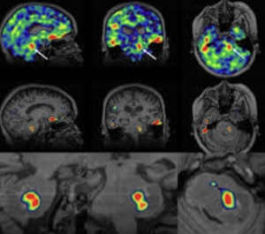
Sodium MRI in Pediatric Brain Tumors

In a narrative review Bhatia et al. from the Children’s Hospital of Philadelphia, Radiological Sciences Laboratory, School of Medicine, Stanford University, published in the American Journal of Neuroradiology to explore the potential of sodium-23 MRI (^23Na-MRI) as a noninvasive imaging modality to assess physiological and biochemical changes in pediatric brain tumors and concluded that is a promising, noninvasive imaging modality capable of providing unique physiological and biochemical information that is not accessible through conventional MRI techniques

This narrative review attempts to position ^23Na-MRI as a frontier imaging technique for pediatric brain tumors. It lauds the modality’s potential to reveal sodium-dependent physiological alterations — but quickly devolves into technological evangelism with minimal clinical anchoring. The piece is high on optimism, low on pragmatism, and entirely devoid of data-supported clinical outcomes.

The article enthusiastically describes the theoretical virtues of sodium MRI — sensitivity to cell integrity, ionic gradients, extracellular space — but offers no compelling clinical cases, no comparative metrics, and no outcome data. What remains is a speculative wish list, presented as a roadmap. The authors confuse imaging potential with diagnostic utility, a common pitfall in radiology reviews driven by physics rather than patient care.

“Exciting” is not a scientific category.

Read more

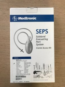
Effectiveness of subdural evacuating port system (SEPS) and middle meningeal artery embolization (MMAE) for chronic subdural hematomas – a multicenter experience

Pairing two well-known procedures—SEPS and MMAE—does not inherently create innovation. Yet, the authors present this as a groundbreaking paradigm, despite:

  • No control group (e.g., SEPS alone, MMAE alone),
  • No randomization,
  • No comparative outcome measures beyond radiographic volume.

It’s procedural layering disguised as progress.

Read more