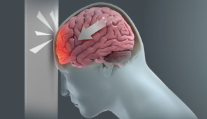
In a retrospective cohort review, Freeman et al. from the University of Colorado, Aurora published in the Journal of Neurosurgery analyzed the sensitivity and specificity of the modified Brain Injury Guidelines (mBIG)—especially mBIG 3 criteria—to predict neurosurgical intervention, and explored the predictive value of individual radiographic parameters.
→ mBIG 3 criteria showed 99.5% sensitivity, and combined mBIG 2+3 reached 100% sensitivity. → Specificity remains low:
-
mBIG 3: 37.2%
-
mBIG 2+3: 18.1%
→ Isolated IPH or SAH in mBIG 3 with GCS 13–15 are poor predictors of intervention. → Authors propose eliminating routine repeat head CT in mBIG 1–2 cases.
🧠 Critical Review
➤ Strengths:
-
Large sample (n = 1128) over 3.5 years (May 2020–Dec 2023).
-
Addresses key clinical issue: reducing unnecessary repeat CTs.
-
High sensitivity makes mBIG a safe exclusion tool, especially mBIG 2+3.
➤ Limitations:
-
Retrospective design → risk of selection bias and unmeasured confounding.
-
Low specificity → risk of overtriage, especially in mBIG 3.
-
Single-center → limits external generalizability.
-
Sparse detail on intervention timing and type.
-
No external validation; subgroup analyses were post hoc.
➤ Interpretation:
-
Excellent rule-out utility — captures nearly all patients needing neurosurgical care.
-
Poor rule-in capacity — high false positive rate may increase resource use.
-
Radiographic IPH/SAH alone, in GCS 13–15 cases, not reliable predictors of need for surgery.
✅ Verdict & Takeaway
Score: '7.0 / 10‘ → Strong cohort and relevant clinical insight. → Undermined by retrospective nature, low specificity, and lack of external validation.
Bottom Line for Neurosurgeons: Use mBIG as a reliable safety net to rule out cases unlikely to require neurosurgical intervention. However, in mild TBI with isolated IPH or SAH, conservative observation without early repeat CT may be acceptable — despite mBIG 3 classification.